What is MEA?
Microelectrode arrays, also known as multielectrode arrays, measure extracellular electrical activity. When excitable cells like neurons or cardiomyocytes are cultured on an MEA, microelectrodes detect their firing in real time. Noninvasive measurements monitor activity for the entire culture duration making MEA an ideal instrument to:
>> Characterize your cells
>> Study development
>> Investigate disease progression
>> Evaluate toxicity testing
Transcript of What is MEA video
Understanding how a cell functions is critical to appreciating its role in the body and disease.
The ability to transmit electrical signals is a key function of many cells. Historically, studying electrical signals required difficult techniques, like manual patch clamp, requiring many months of training to study one cell at a time.
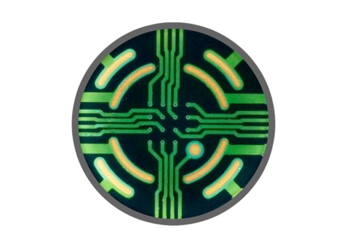
In contrast, MEA, which stands for microelectrode array, also known as multielectrode array, effortlessly and comprehensively measures electrical signals from many cells. An MEA is a grid of small electrodes. When embedded in the surface of a well, and using only basic cell culture techniques, researchers plate electrically active cells on top of the electrodes and measure their activity without disrupting the cells.
While there are many types of electrically active cells in the human body, including: heart cells, retina, muscle, and more, this video will focus specifically on neurons.
Neurons form spontaneous networks as they branch out and communicate across synapses. MEA electrodes are sensitive enough to detect the activity of individual neurons. The signal shown here represents the voltage over time from a single electrode. Each time the neuron above fires an action potential, a spike appears in the signal. Spikes are simultaneously detected from multiple electrodes across the array, making it ideal to monitor activity throughout the culture and investigate network behavior.
A simple way to visualize this neural network behavior is with a raster plot. Each tick mark represents the time, on the x axis, that a spike occurred. Each row of tick marks represents the spikes from a single electrode. A well-wide raster plot shows the activity from all of the electrodes in the well, providing important insights into the behavior of the culture.
Do the neurons fire randomly? Do they fire regularly with a high level of coordination? Are the bursts short? Or long? By looking at different metrics that describe the firing patterns, such as activity, synchrony, and network oscillations, an activity profile of the culture is made. By looking at different activity measures—like how active the culture is, how coordinated the activity is, and the patterns of neural oscillation—an activity profile of the culture is made. These activity profiles act as a fingerprint for the culture, changing based on the composition of cells in culture, their health, maturity, genetic makeup, or chemical effects.
With the Maestro multiwell MEA, neural activity can be monitored from multiple cultures in up to 96 wells at the same time. Because the measurement is noninvasive, the activity can be measured from the same plates many times over months.
With MEA, any cell culture lab can easily conduct important electrophysiological experiments. Whether characterizing an iPSC-derived culture, studying development, disease progression, or screening compounds for safety and efficacy, the detailed functional data from MEA provides critical information.
How could Maestro MEA help in your research?
Visit our Coffee Break Webinars webpage to learn how other researchers are using Maestro MEA assays to enhance their research, or explore the other capabilities of Maestro’s bioelectronic assays at axionbio.com.