What if you could scale your TEER assay without adding more hands-on work?
Transepithelial electrical resistance (TEER) provides a noninvasive way to quantify cellular barrier integrity over time. Traditional TEER methods, however, require researchers to manually position chopstick-style electrodes in each well. As experiments grow, this hands-on process becomes increasingly time-consuming and can introduce variability through differences in electrode placement, timing, and environmental conditions.
Integrated electrodes automate TEER measurements across multiple wells, making it possible to compare more conditions and replicates while monitoring barrier function continuously and consistently over time.
In this video, learn what TEER measures, how electrical resistance relates to barrier integrity, how traditional and integrated-electrode TEER methods compare, why cell confluence matters, and how TEER assays can be used to study barrier function in disease modeling and drug development.
What will you learn?
- What TEER is and how it measures cellular barrier integrity
- How TEER values relate to barrier function
- How traditional manual TEER measurements are performed and how integrated electrodes reduce hands-on work
- Why cell confluence is important when interpreting TEER
- How TEER and confluence can be monitored simultaneously
- How TEER can measure functional responses such as CFTR-mediated ion transport
What is TEER?
TEER stands for transepithelial electrical resistance. For endothelial models, it may also be described as transendothelial electrical resistance. It is an electrical measurement used to quantify the integrity of cellular barriers formed by epithelial or endothelial cells.
Epithelial tissues form barriers in organs including the skin, lungs, liver, kidneys, and digestive tract, while endothelial cells form the inner lining of blood vessels. These cells form tightly packed layers connected by junction proteins, creating selectively permeable barriers that regulate movement between biological compartments.
TEER provides a way to measure the integrity of these barriers in vitro without disrupting the cell culture.
How does TEER measure barrier integrity?
TEER measures how strongly a cell layer resists the flow of electrical current.
Traditionally, TEER is measured by placing two electrodes on opposite sides of a confluent cell layer and applying a low-frequency electrical current. The resistance encountered by the current provides information about the integrity of the cellular barrier.
When cells form a strong, tightly connected barrier, electrical resistance is higher. If the barrier is weak or disrupted, current can flow more easily between the cells, resulting in a lower TEER measurement.
Because the measurement is noninvasive, TEER can be used to follow changes in barrier function over time.
Challenges with traditional TEER measurements
Traditional TEER measurements commonly use manually positioned, chopstick-style electrodes. While this approach can quantify barrier integrity, several experimental factors can introduce variability.
Electrode placement can differ between measurements, environmental conditions can change while cultures are being handled, and collecting measurements manually can limit throughput.
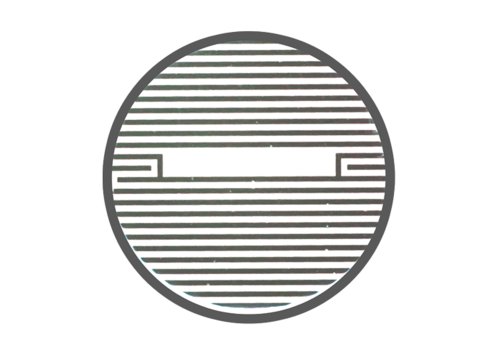
Integrating electrodes directly into the culture well provides an alternative approach. Fixed electrode placement can improve measurement consistency while enabling measurements to be collected automatically across multiple wells without repeatedly disturbing the culture.
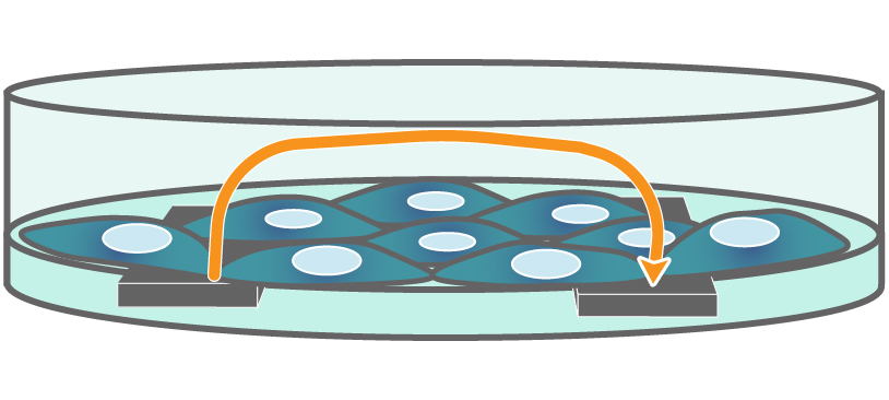
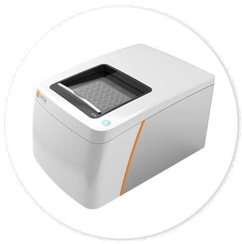
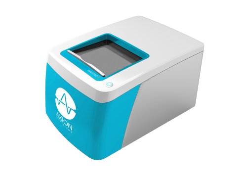
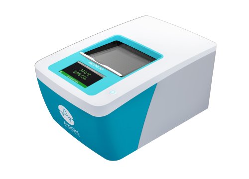
Maestro Z systems use integrated electrodes to automate TEER measurements in a controlled environment, with Maestro ZHT supporting simultaneous monitoring of up to 384 wells. Barrier function can be followed continuously over minutes, hours, or days.
Why does cell confluence matter when measuring TEER?
A fully confluent cell layer is necessary for accurately assessing barrier function with TEER. However, confluence and barrier integrity do not measure the same biological property.
Confluence describes how completely cells cover the culture surface, while TEER reflects the resistance produced by the cellular barrier.
This distinction can be seen when comparing Calu-3 and A549 epithelial cell lines. In the example presented in the webinar, both cell lines reached full coverage within 24 hours. However, only Calu-3 cells expressed tight junction proteins that produced a strong barrier. Their TEER stabilized after the cells had reached confluence.
A culture can therefore reach full coverage without necessarily forming a strong functional barrier.
Measuring TEER and confluence simultaneously
Measuring TEER alongside confluence can provide additional context for interpreting changes in barrier function.
The Maestro Z uses measurements at multiple electrical frequencies to distinguish between the two. Low-frequency measurements are sensitive to barrier properties, while high-frequency measurements detect cell confluence across the bottom of the well.
Monitoring both measurements helps researchers determine whether changes in TEER reflect barrier function rather than differences in cell coverage.
It can also help identify when a culture has reached confluence and when its barrier function has subsequently stabilized.
Using TEER to study CFTR function
TEER can also be used to investigate functional changes in epithelial barriers associated with specific biological mechanisms.
The cystic fibrosis transmembrane conductance regulator (CFTR) is an anion channel involved in regulating secretion and absorption across multiple tissues. CFTR function is particularly important in cystic fibrosis, where disrupted ion transport contributes to altered mucus secretion.
In the example presented in the webinar, stimulation of CFTR causes the channel to open and enables ion transport across the cell membrane. This produces a rapid, substantial decrease in TEER.
When a selective CFTR inhibitor is introduced, the decrease in TEER is blocked in a dose-dependent manner, confirming CFTR as the mechanism underlying the response.
This demonstrates how TEER can measure not only the state of a cellular barrier, but also dynamic functional responses that alter ion transport across that barrier.
TEER assays for disease modeling and drug development
Changes in epithelial and endothelial barrier integrity are relevant to many biological processes and disease states. Because TEER provides a quantitative, noninvasive measurement of barrier function, researchers can monitor cellular barriers over time and evaluate how they respond to disease-related changes, compounds, or other experimental conditions.
TEER can be applied across a variety of epithelial and endothelial barrier models, including:
- Blood-brain barrier models
- Gastrointestinal models
- Pulmonary models
- Organ-on-a-chip models
Across these models, researchers can use TEER to investigate how barrier integrity develops, stabilizes, or becomes disrupted. These measurements can also support studies evaluating drug or chemical transport and the effects of potential therapeutic compounds on barrier function.
For disease modeling and drug development, longitudinal TEER measurements provide functional information about an barrier without requiring researchers to disrupt the culture at each timepoint. When measured alongside cell confluence, TEER can also help distinguish changes in barrier function from changes in cell coverage.
Explore TEER and barrier function assays to learn more about applications for monitoring epithelial and endothelial barriers in vitro.

“The Teer assay is a game-changer in cell barrier research.”
The Teer assay is a vital tool for unraveling the mysteries of cellular barriers and advancing our understanding of health and disease in the easiest way possible. We utilize the Maestro Edge system with the Impedance Module to measure TEER in colonic epithelial cells cultured with various bacteria in vitro. Recently, we demonstrated that B. adolescentis bacteria impair epithelial barrier integrity in patients with irritable bowel syndrome (IBS).
- Hadar Bootz-Maoz. Bar Ilan University, Ramat Gan, Israel