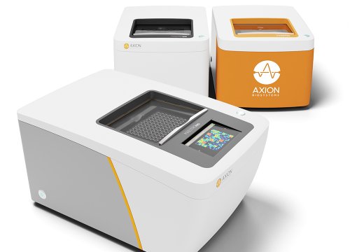
Evaluate proarrhythmic risk with human iPSC-derived cardiomyocytes and scalable, label-free functional electrophysiology.
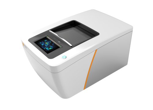
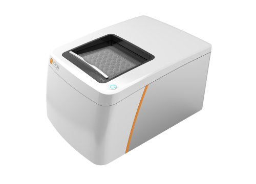
The Maestro MEA platform measures integrated cardiac function—including rhythm, repolarization, conduction, contractility, and action-potential morphology—to help researchers identify potential cardiac liabilities earlier in drug development.
Human iPSC-cardiomyocyte MEA assays are also among the most mature examples of stem cell-derived New Approach Methodologies (NAMs) advancing toward broader pharmaceutical and regulatory use.
Human-relevant functional cardiac assessment
MEA technology provides a scalable, human-relevant approach for evaluating cardiac electrophysiology in vitro.
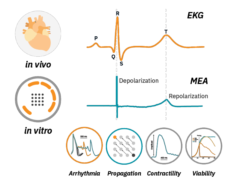
Analogous to an in vivo ECG, MEA field potentials capture changes in depolarization, repolarization, rhythm, and beat timing across a cardiomyocyte syncytium.
The Maestro MEA platform adds complementary functional readouts:
- Arrhythmia detection
Detect rhythm abnormalities and action-potential morphology with LEAP. - Propagation and conduction
Measure slowed or disrupted electrical conduction across the cardiac network. - Contractility assessment
Evaluate electrical and mechanical function together. - Viability multiplexing
Distinguish functional effects from broader changes in cell health.
Real-time, label-free in vitro cardiac analysis
-
Key evidence supporting human iPSC-cardiomyocyte MEA for cardiac safety>
-
Measure integrated cardiac electrophysiology in real time>
-
Add action-potential morphology without giving up MEA scale>
-
Establish functional acceptance criteria for hiPSC-cardiomyocyte assays>
-
Track conduction across the cardiac syncytium>
-
Elicit mature force-frequency responses with chronic pacing>
The cardiac MEA assay has been evaluated through international, multi-site studies designed to assess assay reproducibility and prediction of proarrhythmic risk.
In a blinded multi-site study described by Millard et al. (2018), eight compounds were evaluated in 18 studies across 13 global sites, three MEA platforms, and four hiPSC-cardiomyocyte sources.
For Maestro MEA:
- 85% of participating sites passed initial quality-control criteria.
- Test compounds were correctly identified 93% of the time based on the defined field-potential response.
- Baseline well-to-well FPDc variability was 6.4%.
- Maestro MEA demonstrated the lowest well-to-well variability among the evaluated platforms.
| Maestro MEA | MEA2100 | xCELLigence RTCA CardioECR |
| Most Reliable Sites that passed QC | 85% | 42% | 25% |
| Most Accurate Identified compounds expected to induce a 20% change in FPDc | 93% | 50% | 75% |
| Lowest Variability Well-to-well FPDc variability in baseline | 6.4% | 21.5% | 15.2% |
A subsequent CiPA study expanded testing to 28 blinded compounds representing low, intermediate, and high proarrhythmic risk and demonstrated strong agreement between study sites.
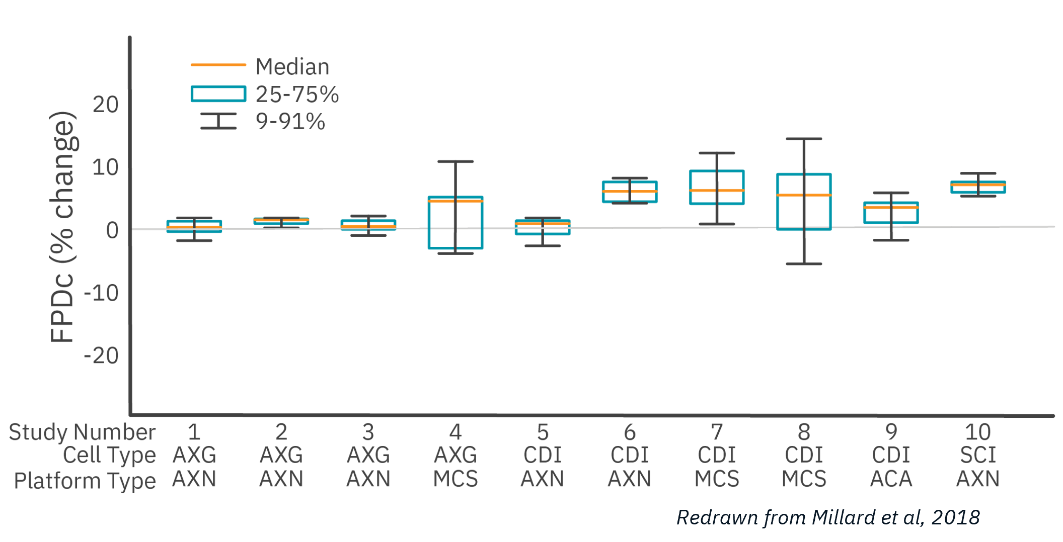
Of the 10 studies that passed QC acceptance criteria, Maestro MEA had the lowest well-to-well variability, 58% less than the next lowest despite being performed across 6X as many sites.
The Maestro MEA assay correctly identified test compounds 93% of the time. A follow-up study (Blinova et al 2018) expanded the scope to 28 blinded compounds of low, intermediate, and high risk. Maestro MEA had a 0.93 correlation coefficient between study sites.
Together, these studies illustrate an important requirement for NAM adoption: human-relevant biology must be paired with reproducible, standardized measurement.
Cardiomyocytes cultured on an MEA form a spontaneously beating, electrically connected syncytium.
As action potentials propagate across the culture, electrodes record extracellular field potentials that reflect key features of cardiac electrical activity.
MEA recordings can quantify:
- beat period and beat rate;
- depolarization amplitude;
- field potential duration;
- repolarization timing;
- rhythm irregularity;
- arrhythmic events;
- conduction velocity;
- propagation pattern.
Because recording is label-free and noninvasive, researchers can collect baseline and post-treatment measurements from the same cultures.
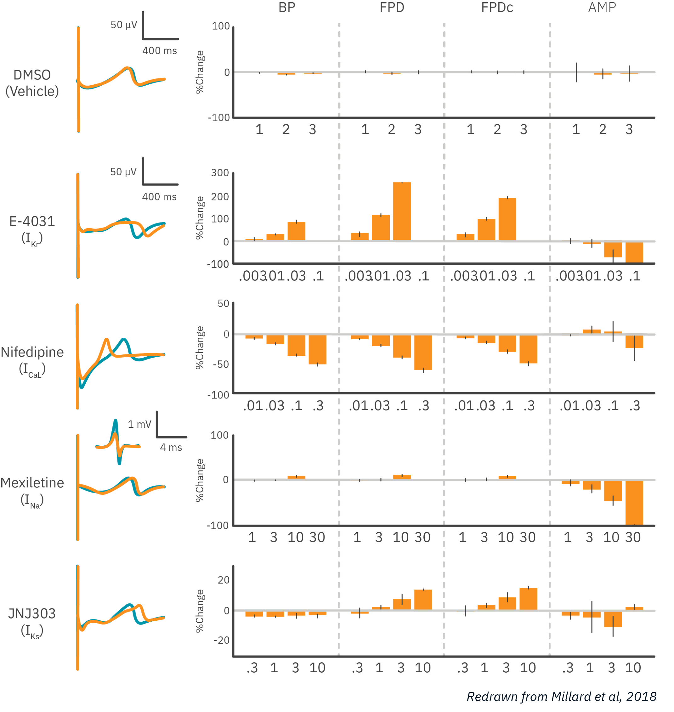
Compound effects demonstrated with MEA
Human iPSC-derived cardiomyocytes were treated with compounds targeting major cardiac ion channels.
The MEA assay detected the expected dose-dependent electrophysiological responses:
- potassium-channel block prolonged field potential duration;
- calcium-channel block shortened field potential duration;
- sodium-channel block reduced the amplitude of depolarization.
These functional responses demonstrate how MEA can distinguish mechanisms affecting different components of cardiac electrophysiology.
Field potentials provide a scalable measurement of cardiac electrophysiology, but action-potential morphology can reveal additional mechanistic information about changes in depolarization, plateau, and repolarization.
LEAP (local extracellular action potential), is an Axion MEA recording capability that captures action-potential-like waveforms from intact cardiomyocyte monolayers.
LEAP adds waveform-level information while maintaining the advantages of the existing MEA workflow:
- multiwell scalability;
- label-free recording;
- intact cardiomyocyte syncytia;
- software-guided induction;
- the same MEA plates and cell culture;
- integration with standard field-potential measurements.
This enables researchers to add deeper electrophysiological information without moving the experiment to a separate single-cell recording platform.
What can LEAP measure?
LEAP provides quantitative endpoints associated with different phases of the cardiac action potential.
Depolarization: Rise time can provide information about effects on the rapid depolarization phase of the waveform.
Repolarization: APD30, APD50, and APD90 quantify action-potential duration at different stages of repolarization.
Action-potential shape: Triangulation identifies changes in repolarization morphology that may provide information beyond action-potential duration alone.
Arrhythmic events: LEAP can support detection of waveform abnormalities including early afterdepolarizations (EADs).
Together, these measurements enable researchers to evaluate not only whether the duration of an action potential changes, but how its morphology changes.
Learn About the FDA iSTAND Program and Axion's Cardiac MEA Assay
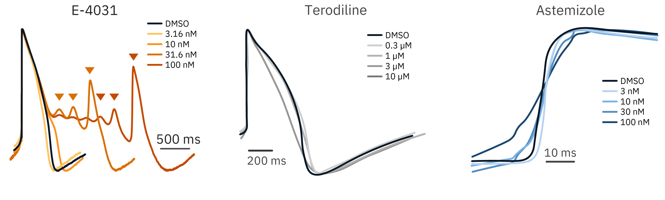
LEAP reveals mechanism-specific changes in action-potential morphology
Compounds affecting different cardiac ion channels can alter distinct regions of the action-potential waveform.
In pharmacology studies presented by Axion, LEAP captured expected changes associated with different electrophysiological mechanisms.
Calcium-channel inhibition
L-type calcium-channel inhibition produced dose-dependent shortening of the LEAP waveform, reflected in reductions in APD30, APD50, and APD90.
hERG-channel inhibition
hERG-channel block prolonged repolarization. At higher concentrations, LEAP recordings also revealed early afterdepolarizations.
Importantly, the prolongation was most evident later in repolarization, demonstrating how measurements at different action-potential durations can help localize waveform changes.
Action-potential triangulation
LEAP can also distinguish changes in waveform shape from changes in duration alone.
Compounds may produce similar overall prolongation while exhibiting substantially different repolarization morphology. Quantifying triangulation and other shape-based endpoints can therefore provide additional information relevant to arrhythmic risk assessment.
Why waveform morphology matters
A measurement of duration tells researchers how long the action potential lasts.
Waveform morphology provides additional information about how the action potential changed.
Bringing these endpoints into a scalable MEA workflow can help translate detailed electrophysiological measurements into larger drug-screening experiments.
Watch: LEAP—Scalable Cardiac Action-Potential Electrophysiology
Biological complexity alone does not make an assay reliable.
For stem cell-derived models to support routine drug-development decisions, researchers must also know that the cells have developed the functional properties required for the assay.
Axion iPSC Model Standards, or AIMS, provide proposed functional criteria for evaluating hiPSC-cardiomyocyte performance on MEA.
These criteria consider measures such as:
- spontaneous beat rate;
- depolarization spike amplitude;
- field potential duration;
- synchronization of activity across the cardiac syncytium.
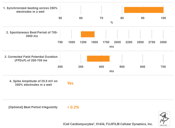
Functional acceptance criteria can help laboratories identify whether a culture is suitable for testing before interpreting compound responses.
This type of standardization is an important step in moving advanced cell models from promising biology toward reproducible NAM workflows.
Cardiac safety workflow

A typical human iPSC-cardiomyocyte MEA safety assay follows a straightforward workflow:
- Plate hiPSC-derived cardiomyocytes on a multiwell MEA plate.
- Culture the cells until stable functional activity is established.
- Confirm baseline activity and assay acceptance criteria.
- Record baseline electrophysiology.
- Add the test compound.
- Allow the culture to equilibrate.
- Record post-treatment field potentials and cardiac metrics.
- Add LEAP measurements when action-potential morphology is required.
- Compare treatment effects across concentrations and controls.
Because the recording is noninvasive, several measurements can be obtained from the same cardiac culture.
Cardiac safety is not determined solely by the behavior of individual cells. Electrical signals must propagate consistently across cardiac tissue. Disruptions in conduction can contribute to re-entrant activity and arrhythmic risk.
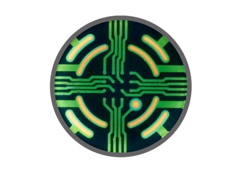
Multiple electrodes distributed across each MEA well allow researchers to measure electrical activity at different locations within the cardiomyocyte syncytium and calculate:
- conduction velocity;
- propagation direction;
- spatial conduction patterns;
- disruption or inconsistency in propagation.
In the example shown on this page, sodium-channel inhibition reduced conduction velocity across the cardiomyocyte network.
A separate anticancer compound disrupted the pattern and consistency of electrical propagation across the syncytium.
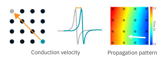
To demonstrate the Maestro MEA’s propagation assay, human iPSC-derived cardiomyocytes were dosed with cardioactive compounds.
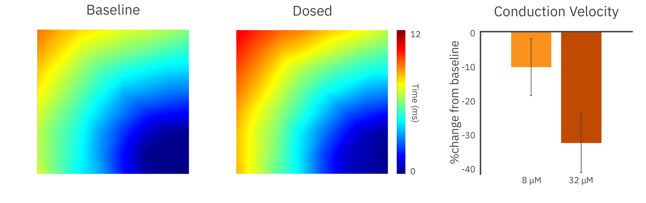
In the presence of a sodium channel blocker, the cardiomyocytes showed a decrease in conduction velocity across the array.
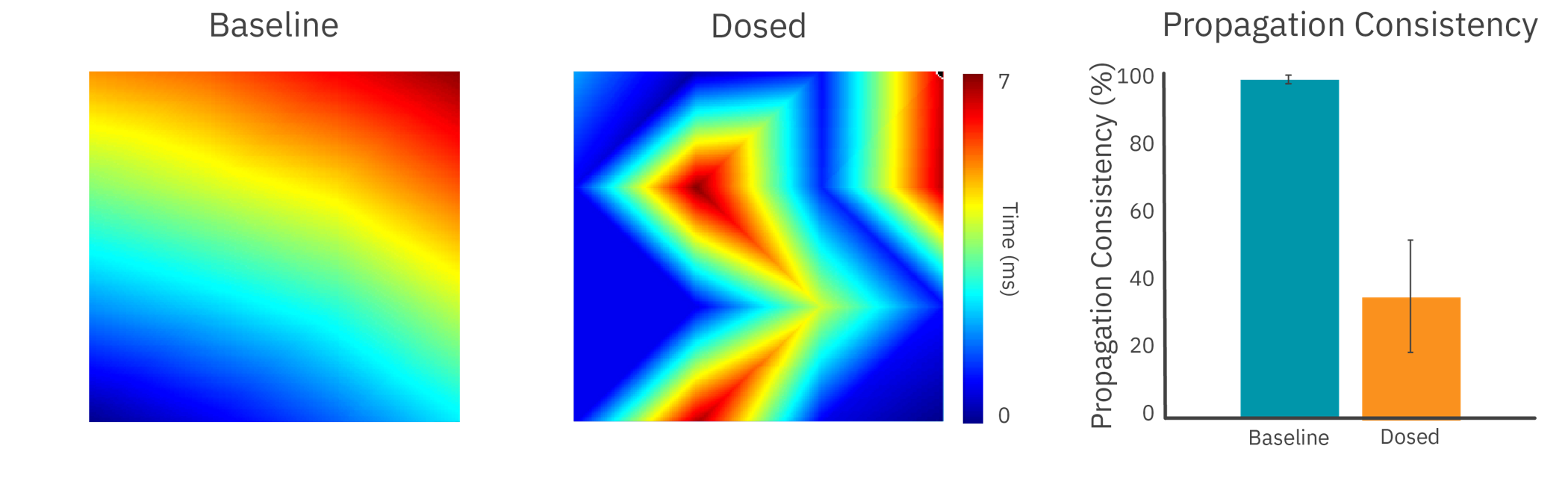
Human iPSC-derived cardiomyocytes were monitored across multiple electrodes to measure electrical propagation. Sodium-channel inhibition reduced conduction velocity, while exposure to an anticancer compound disrupted propagation patterns and reduced conduction consistency across the cardiac syncytium.
Purpose: To elicit a mature force-frequency relationship in hiPSC-derived cardiomyocytes. In a mature cardiomyocyte, an increase in frequency is accompanied by an increase in contractile strength. This force-frequency relationship is often lacking in hiPSC-derived cardiomyocytes and is a sign of their immaturity.
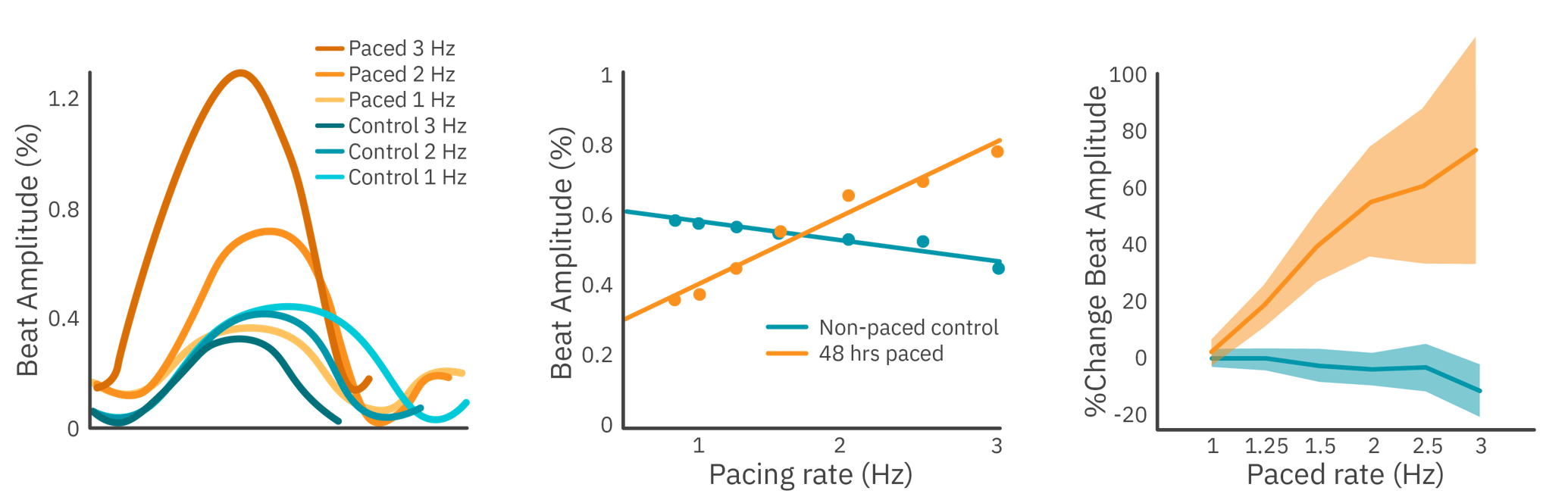
Electrical stimulation was used to measure the force-frequency relationship of hiPSC-derived cardiomyocytes with the Maestro MEA. Prior to testing, the culture was kept on the Maestro MEA for 48 hours, chronically pacing half of the wells.
Results: Cardiomyocytes chronically paced for 2 hours demonstrated a mature force-frequency relationship as measured by beat amplitude using Maestro contractility.
Want to learn how?
From CiPA research to regulatory-facing use
The Comprehensive in vitro Proarrhythmia Assay (CiPA) initiative was established to improve prediction of Torsades de Pointes risk beyond reliance on hERG block and QT prolongation alone.
CiPA Framework
CiPA brought together regulators, industry, and academia around four complementary approaches:
Multi-ion-channel pharmacology
Characterize drug effects across key cardiac ion channels.
In silico human ventricular models
Integrate ion-channel data to model human ventricular electrophysiology.
Clinical ECG analysis
Evaluate clinical repolarization and proarrhythmic risk.
Human iPSC-derived cardiomyocyte assays
Measure integrated functional electrophysiology in human cardiac cells.
Regulatory engagement continues to advance
Human iPSC-cardiomyocyte data are increasingly used as supporting evidence in cardiac safety assessment.
In December 2025, FDA CDER accepted Axion BioSystems’ letter of intent for the Human iPSC-Cardiomyocyte MEA Assay for Prediction of Clinical Cardiovascular Repolarization Risk into the FDA ISTAND program.
Acceptance begins a structured qualification pathway for the assay.
The milestone advances stem cell-derived functional assays as regulatory-facing NAMs.
CiPA research
Foundational science and method development.
Multi-site validation
Cross-lab studies establish reproducibility.
Supporting evidence
Human iPSC-MEA data inform cardiac safety assessment.
FDA ISTAND LOI accepted
December 2025 milestone begins qualification pathway.
Adoption depends on human-relevant biology, reproducibility, functional standards, validation, and a clearly defined context of use.
“The Maestro Pro MEA enables our lab to dramatically scale up our studies focusing on drug-induced cardiotoxicity.”
“Our team utilizes human induced pluripotent stem cell (hiPSC)-derived cardiomyocytes to evaluate cardiotoxic chemotherapeutic agents (cancer drugs that do damage to the heart) and develop safer versions of known cardiotoxic compounds. For pre-clinical drug discovery and screening, the Maestro Pro fits perfectly into our workflows to generate vast amounts of accurate, informative data. We are also excited to explore new possibilities in analyzing more advanced 3D hiPSC-derived cardiac organoids on the Maestro Pro MEA.”
- Arun Sharma, PhD, Cedars-Sinai Medical Center

Featured resources for cardiac safety and cardiotoxicity
Explore validation studies, regulatory developments, application data, protocols, and educational resources supporting human-relevant cardiac safety assessment.
From Stem Cell Models to Trusted NAMs: How Functional Assays Support Drug Development Decisions
Webinar
Learn how human stem cell-derived assays progress from promising biological models to reproducible, standardized NAMs capable of supporting pharmaceutical and regulatory decision-making.
The NAMs Inflection Point: Building a Future-Proof Safety Strategy with Maestro MEA
Webinar
Explore the evolution of human iPSC-cardiomyocyte MEA from CiPA studies to regulatory-facing use, including Axion's participation in the FDA ISTAND pathway.
LEAP: Scalable Cardiac Action-Potential Electrophysiology
Webinar
See how LEAP brings action-potential morphology—including APD, triangulation, and EAD analysis—into a scalable multiwell MEA workflow.
CiPA: Cardiac Safety Studies
Application Note
Review how human stem cell-derived cardiomyocytes and MEA can provide an integrated assessment of compound effects beyond hERG inhibition alone.
Cardiac Metric Definitions
Technical Resource
Explore electrophysiological, propagation, and cardiac-function metrics available for Maestro MEA analysis.
Cardiomyocyte Culture and Assay Protocols
Protocols
Access model-specific guidance for preparing human stem cell-derived cardiomyocytes for cardiac MEA experiments.
Looking for additional cardiac safety studies?
Frequently asked questions about cardiotoxicity MEA assays
A NAM is an approach that provides information relevant to hazard or risk assessment while reducing reliance on vertebrate animal testing.
For cardiac safety, human iPSC-derived cardiomyocyte assays can provide functional information about drug effects in a human cell system.
The value of a NAM depends not only on its biological relevance but also on whether the assay is reproducible, standardized, fit for purpose, and capable of supporting meaningful drug-development decisions.
Human iPSC-derived cardiomyocytes express multiple interacting cardiac ion channels and form electrically active networks in culture.
This enables researchers to study integrated electrophysiological responses rather than assessing one ion channel in isolation.
MEA records extracellular electrical signals generated as cardiomyocytes depolarize, repolarize, beat, and propagate electrical activity across the culture.
Key measurements include beat rate, field potential duration, rhythm, depolarization amplitude, conduction, and arrhythmic events.
Human iPSC-derived cardiomyocytes are used in the CiPA framework as an integrated cellular system for assessing whether predicted ion-channel effects result in functional electrophysiological changes in human cardiac cells.
MEA has been used extensively to record these responses.
Not yet.
In December 2025, FDA CDER accepted Axion BioSystems' letter of intent for the Human iPSC-Cardiomyocyte MEA Assay for Prediction of Clinical Cardiovascular Repolarization Risk into the ISTAND program.
Acceptance begins a structured qualification process; it should not be interpreted as completed FDA qualification.
LEAP stands for Local Extracellular Action Potential.
It is an Axion MEA capability that captures action-potential-like waveforms from intact cardiomyocyte monolayers and enables measurements including APD30, APD50, APD90, rise time, triangulation, and early afterdepolarizations.
No.
LEAP adds an additional waveform-level readout to the MEA workflow. Researchers can continue to collect standard field-potential measurements and add LEAP when more detailed action-potential morphology is useful.
The Maestro MEA platform can measure multiple functional endpoints relevant to cardiac safety, including rhythm, repolarization, action-potential morphology, propagation, and contractility.
Rhythm
Measures: Beat rate, beat period, rhythm regularity
Can reveal: Chronotropic effects and arrhythmia-like behavior
Repolarization
Measures: Field potential duration and corrected FPD
Can reveal: Delayed or shortened repolarization
Action-potential morphology
Measures: APD30, APD50, APD90, rise time, triangulation, EADs
Can reveal: Mechanism-specific waveform changes and electrophysiological instability
Propagation
Measures: Conduction velocity and propagation pattern
Can reveal: Slowed or disrupted conduction across the cardiomyocyte syncytium
Contractility
Measures: Contraction amplitude and excitation-contraction relationships
Can reveal: Changes in mechanical cardiac function