Assessing proarrhythmic risk with hiPSC-cardiomyocytes
hERG inhibition does not tell the whole cardiac safety story. Because compounds can affect multiple cardiac ion channels, their integrated effects on depolarization, repolarization, and rhythm may differ from what a single-channel assay predicts.
The Comprehensive in vitro Proarrhythmia Assay (CiPA) initiative was established to advance how drug-induced arrhythmia risk is evaluated. As part of this effort, the CiPA myocyte studies investigated the use of human stem cell-derived cardiomyocytes and microelectrode array (MEA) technology to characterize functional cardiac responses to compounds in vitro.
In this video, learn why the CiPA initiative was developed, how cardiomyocyte MEA assays measure drug-induced changes in cardiac electrophysiology, and how different ion channel effects can be distinguished using functional measurements.
What will you learn?
- Why drug-induced arrhythmia is an important consideration in cardiac safety assessment
- How hERG block and QT prolongation have traditionally been used to identify cardiac risk
- Why hERG inhibition alone does not always predict proarrhythmic risk
- How the CiPA initiative proposed advancing cardiac safety assessment
- How cardiomyocyte MEA assays measure depolarization and repolarization
- How different ion channel blockers produce distinct electrophysiological responses
Why was the CiPA initiative developed?
Drug-induced changes in cardiac electrophysiology can lead to potentially serious arrhythmias and have contributed to late-stage drug development failures and market withdrawals.
One important cardiac safety concern is torsades de pointes (TdP), a potentially fatal ventricular arrhythmia associated with delayed cardiac repolarization. Delayed repolarization can manifest clinically as prolongation of the QT interval on an electrocardiogram (ECG).
Many drugs associated with delayed repolarization and TdP have also been shown to block the hERG potassium current. As a result, cardiac safety screening has historically placed substantial emphasis on hERG channel assays and in vivo QT measurements.
These approaches have helped prevent drugs with proarrhythmic liabilities from advancing, but hERG inhibition does not always translate directly into clinical arrhythmia risk.
Why isn't hERG block alone enough to assess proarrhythmic risk?
Because a compound can affect multiple cardiac ion channels, measuring its interaction with only one channel may not provide a complete picture of its functional cardiac effects.
The video highlights verapamil as an example. Although verapamil can potently block the hERG potassium current, its activity as an L-type calcium channel blocker counterbalances this effect, and it does not produce the QT prolongation or torsades de pointes risk that might be expected from hERG inhibition alone.
Examples like this helped motivate efforts to reconsider how drug-induced arrhythmia risk is evaluated and to incorporate methods capable of measuring more integrated cardiac responses.
What is the CiPA myocyte assay?
The CiPA myocyte assay uses beating human induced pluripotent stem cell-derived cardiomyocytes (hiPSC-CMs) as an in vitro model for evaluating drug-induced changes in cardiac electrophysiology.
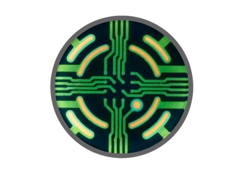
Microelectrode array technology records the extracellular cardiac field potential generated as cardiac action potentials propagate across a functionally connected cardiomyocyte network.
This field potential provides measurable features associated with cardiac depolarization and repolarization, conceptually similar to how an ECG reflects electrical activity propagating through the heart.
From these recordings, researchers can quantify functional characteristics, including:
- Beat frequency
- Depolarization amplitude
- Repolarization timing
- Repolarization irregularities
Repolarization timing and irregularities are particularly relevant when investigating electrophysiological changes associated with proarrhythmic risk.
How does MEA measure drug-induced cardiac electrophysiology?
Microelectrode array technology enables researchers to noninvasively record extracellular electrical activity from interconnected cardiomyocytes.
Because MEA measurements capture the integrated electrophysiological behavior of the cardiac cell network, researchers can observe how different compounds alter depolarization, repolarization, beat timing, and rhythm.
The earlier CiPA pilot study evaluated endpoints including beat period, field potential duration, spike amplitude, and arrhythmic events across multiple hiPSC-cardiomyocyte types, MEA platforms, and independent test sites.
These measurements provide a functional complement to assays examining individual ion channels.
Distinguishing different ion channel effects with MEA
Different ion channel blockers can produce distinct changes in cardiomyocyte field potentials.
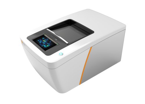
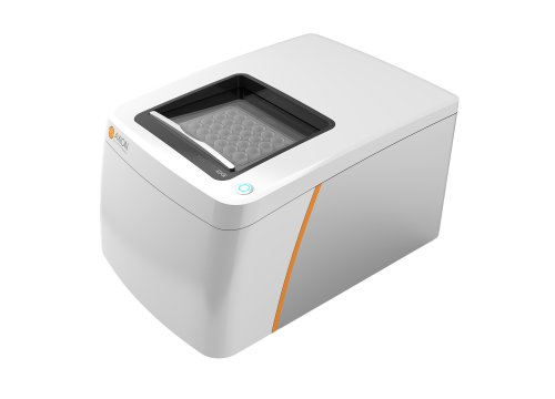
The video demonstrates three examples using Maestro MEA:
Moxifloxacin and hERG potassium channel block
Moxifloxacin, a hERG potassium channel blocker, produced a concentration-dependent prolongation of repolarization along with repolarization irregularities.
This response illustrates how inhibition of the repolarizing potassium current can be detected through changes in the cardiac field potential.
Nifedipine and L-type calcium channel block
In contrast, the L-type calcium channel blocker nifedipine shortened repolarization as concentration increased.
This contrasting response illustrates why integrated electrophysiological measurements can provide information beyond whether a compound interacts with hERG alone.
Mexiletine and sodium channel block
The sodium channel blocker mexiletine reduced depolarization spike amplitude, demonstrating another distinct electrophysiological phenotype measurable using cardiomyocyte MEA recordings.
Together, these examples show how functional MEA measurements can distinguish compound effects associated with different cardiac ion currents.
Evaluating hiPSC-cardiomyocyte assays across sites
For a cardiac safety assay to support broader drug development workflows, its results need to be reproducible across laboratories.
The blinded CiPA MEA pilot study evaluated eight compounds across 18 individual studies using four cardiomyocyte types, three MEA platforms, and 13 performance sites.
The study found that standardized experimental protocols and pharmacological assay qualification improved consistency across sites and supported the continued evaluation of hiPSC-cardiomyocyte assays for cardiac safety assessment.
Read the published CiPA pilot study to explore the experimental design and cross-site results in more detail.
Advancing in vitro cardiac safety assessment with CiPA
The CiPA initiative reflects a shift toward evaluating proarrhythmic risk using multiple sources of mechanistic and functional information.
Human stem cell-derived cardiomyocytes provide a model in which the integrated electrophysiological effects of a compound can be measured directly. When combined with MEA technology, researchers can characterize changes in cardiac depolarization, repolarization, beating, and arrhythmic activity without disrupting the cellular network.
These measurements help distinguish the effects of compounds targeting different cardiac ion channels and support a more comprehensive assessment of potential cardiac liabilities.
Watch the video to learn how the CiPA myocyte assay uses hiPSC-derived cardiomyocytes and MEA technology to investigate drug-induced electrophysiological effects and proarrhythmic risk.
Explore the CiPA research
Continue exploring the CiPA studies and the use of hiPSC-derived cardiomyocytes for cardiac safety assessment:
>> Explore the validation results: See the CiPA validation study results and learn what the multisite study demonstrated about human stem cell-derived cardiomyocytes and assay reproducibility.
>> Explore the MEA assay workflow: Learn about optimizing MEA assays for hiPSC-cardiomyocyte cardiac safety studies, including signal quality, environmental control, time-matched controls, and analysis.
>> Read the research: Explore the published CiPA pilot study examining the cross-site reliability of hiPSC-cardiomyocyte MEA assays.