CiPA Phase II: Optimizing MEA assays for hiPSC-cardiomyocyte cardiac safety studies
The right cell model is only the starting point for a cardiac safety assay. Reproducible results depend on the entire workflow, from signal quality and experimental controls to environmental stability and standardized analysis.
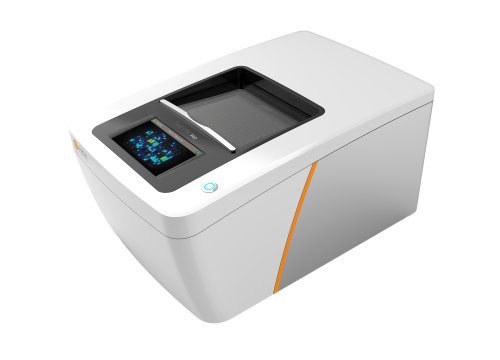
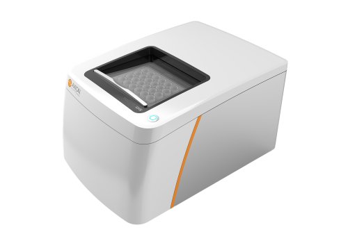
In this video, explore the technical considerations behind using microelectrode array (MEA) technology with human induced pluripotent stem cell-derived cardiomyocytes (hiPSC-CMs) for cardiac safety assessment. Using the CiPA Phase II study as context, learn how the Maestro MEA platform was designed to support accurate measurement of cardiac electrophysiology across multiwell assays.
What will you learn?
- Why signal quality matters when measuring cardiac depolarization and repolarization
- How simultaneous controls support interpretation of compound effects
- Why temperature and CO2 stability are important for cardiomyocyte assays
- How multiwell MEA expands cardiac electrophysiology across conditions and controls
- How software can simplify field potential analysis and arrhythmia classification
- How standardized analysis and reporting can support CiPA workflows
Why does MEA signal quality matter for cardiac safety assays?
Cardiac safety assays using hiPSC-CMs rely on accurate measurement of electrophysiological signals.
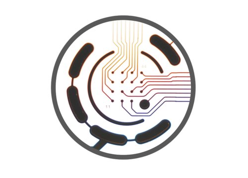
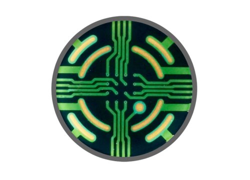
When cardiomyocytes are cultured over MEA electrodes, their electrical activity produces extracellular field potentials. Features within these signals provide information about cardiac depolarization and repolarization, including field potential duration (FPD).
Clear signals make these electrophysiological features easier to identify and quantify. Signal quality is therefore an important consideration when evaluating compound-induced changes in cardiac activity, particularly when those effects are subtle.
The Maestro MEA platform and MEA plates are designed to capture cardiac field potentials with sufficient resolution to identify depolarization and repolarization features and quantify changes following compound exposure.
Why are time-matched controls important?
When studying the effects of a novel compound, researchers need appropriate controls to distinguish treatment-related changes from variability that occurs over the course of an experiment.
Running positive, negative, and vehicle controls alongside experimental conditions on the same plate enables these groups to be evaluated under the same environmental and temporal conditions.
The Maestro MEA system records from 768 electrodes simultaneously across an industry-standard 96-well plate, allowing multiple experimental conditions and time-matched controls to be incorporated into the same assay.
This multiwell approach allows researchers to collect electrophysiological data across conditions in parallel rather than relying on measurements collected at different times.
Maintaining a stable environment for cardiomyocyte recordings
Cardiomyocyte electrophysiology is sensitive to environmental conditions. Changes in factors such as temperature and CO2 can alter beating behavior, potentially introducing variability into an experiment.
Maintaining a stable environment during data collection therefore helps researchers distinguish pharmacological effects from changes caused by experimental conditions.
The Maestro MEA environmental chamber controls temperature and CO2 during recordings while minimizing electrical interference, allowing cardiomyocyte activity to be monitored under stable conditions.
This environmental control can support both short- and long-term measurements without repeatedly moving cultures between an incubator and recording instrument.
Analyzing cardiac electrophysiology in real time
Collecting electrophysiological signals is only one part of a cardiac MEA assay. Researchers also need to identify and quantify relevant changes in cardiomyocyte behavior.
AxIS software enables researchers to set up, run, and analyze MEA experiments while monitoring cardiac activity as data are collected.
For CiPA workflows, the analysis tools described in the video were designed to help researchers:
- Track cardiac repolarization
- Monitor beating characteristics
- Detect and classify arrhythmic events
- Review and verify field potential measurements
- Generate standardized reports for CiPA study data
Providing access to both automated analysis and the underlying electrophysiological signals allows researchers to evaluate detected responses and verify measurements when needed.
Detecting drug-induced changes in cardiac electrophysiology
Functional cardiac safety assays need to capture changes in cardiomyocyte electrophysiology that may indicate potential cardiac liabilities.
MEA recordings can quantify parameters including depolarization amplitude, field potential duration, beat timing, and irregular beating.
Together, these measurements can help researchers characterize how hiPSC-derived cardiomyocytes respond to test compounds and identify electrophysiological changes associated with potential proarrhythmic effects.
The CiPA initiative provided an important framework for evaluating how these human cell-based assays could contribute to preclinical cardiac safety assessment.
Supporting reproducible hiPSC-cardiomyocyte MEA assays
Reliable MEA-based cardiac safety assays depend on the complete experimental workflow rather than a single instrument specification.
Signal quality supports accurate identification of cardiac electrophysiological features. Multiwell recording enables experimental conditions and controls to be measured simultaneously. Environmental regulation helps maintain stable cardiomyocyte behavior, while analysis software provides tools for quantifying electrophysiological changes and identifying arrhythmic events.
Together, these capabilities supported the use of Maestro MEA in the multi-site CiPA studies evaluating hiPSC-derived cardiomyocytes for cardiac safety assessment.
Watch the video to learn how the Maestro MEA platform supports multiwell hiPSC-cardiomyocyte assays and the measurement and analysis of cardiac electrophysiology for CiPA studies.
Explore the CiPA research
The CiPA program evaluated hiPSC-derived cardiomyocytes and MEA technology through multiple stages of assay development and validation.
>> Explore the validation results: Read about the CiPA validation study results and how human iPSC-derived cardiomyocytes were evaluated across independent test sites.
>> Read the research: Explore the published CiPA pilot study examining cross-site reliability of hiPSC-CM cardiac safety assays using MEA technology.