Understand what electrode density changes, what it does not, and which system characteristics matter for your research.
What is the difference between conventional MEA and HD-MEA?
Conventional microelectrode arrays (MEAs), also called standard or classic MEAs, and high-density MEAs (HD-MEAs) both record extracellular electrical activity. Conventional MEA supports established firing, bursting, and network measurements; HD-MEA adds denser spatial sampling that can support more detailed local and unit-level analysis.
Neither is universally better. The right approach depends on the biological question, simultaneous recording capacity, experimental scale, and analysis workflow.
Axion BioSystems manufactures both conventional MEA and HD-MEA technology. Its Synexus platform combines these recording approaches with high-content electrical imaging, giving researchers the option to select established network assays or finer spatial measurements within a configurable multiwell system.
This guide compares what electrode density changes, what it does not, and which system characteristics matter when selecting an MEA platform.
How do conventional MEA and HD-MEA work?
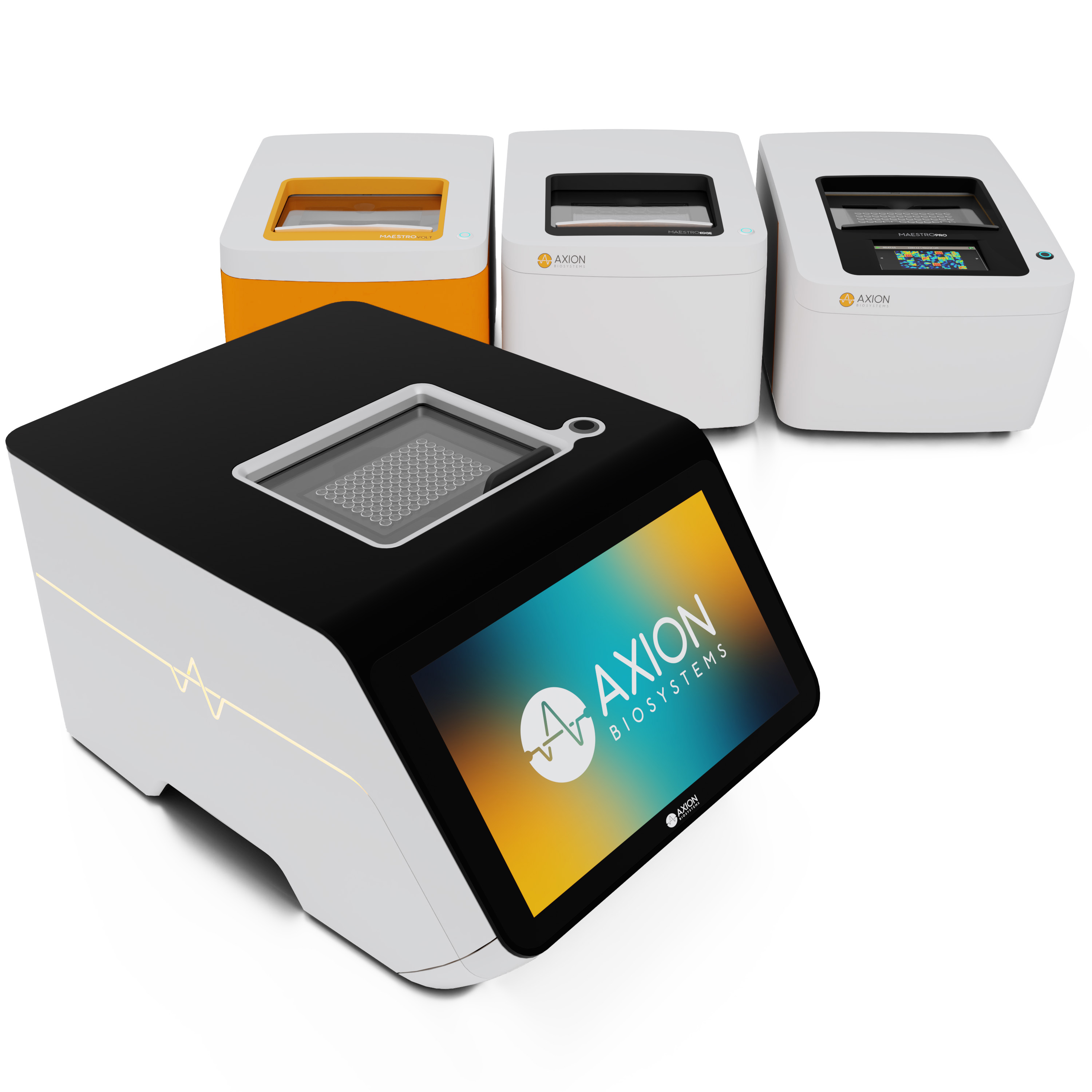
Both technologies measure extracellular voltage changes from electrically active cells cultured on, or positioned against, an electrode array. Their recordings support firing rate, bursts, network bursts, synchrony, longitudinal changes, and responses to compounds or stimulation. HD-MEA uses a more densely sampled array, often implemented in CMOS.
There is no universal electrode-count threshold defining HD-MEA. Recording performance depends on sensing geometry, cell-electrode contact, noise, sampling rate, simultaneous channel capacity, and the analysis required for the endpoint.
Conventional MEA vs. HD-MEA at a glance
| Consideration | Conventional MEA | High-density MEA (HD-MEA) |
| Measurement principle | Extracellular electrical recording | The same extracellular recording principle |
| Spatial sampling | Typically 8-64 electrodes with 200-400 μm electrode spacing | Hundreds to thousands of electrodes under 100 μm spacing. (Synexus has >138k electrodes with 12.5 μm spacing) |
| Common endpoints | Firing, bursting, network coordination, and treatment responses | The same endpoints, with additional spatial and potentially unit-level information |
| Individual-neuron analysis | Possible under suitable conditions, but separation and coverage may be limited | Dense multichannel signals can provide more information for spike sorting and localization |
| Fine spatial measurements | Depends on array layout, signal quality, and sample positioning | Suitable systems can support local circuit and subcellular investigations |
| Longitudinal studies | Repeated population measurements | Repeated measurements |
| Multiwell experiments | Available on appropriate systems | Also available on appropriate systems; simultaneous well throughput depends on the platform |
| Analysis requirements | Many assays use electrode-level and network-level endpoints | Spatial mapping and single-unit analysis can add complexity |
Who manufactures high-density MEA (HD-MEA) systems?
Manufacturers of HD-MEA systems include Axion BioSystems, MaxWell Biosystems, and 3Brain.
>> Axion BioSystems offers Synexus, a configurable platform integrating conventional MEA, HD-MEA, and complementary high-content electrical imaging. Synexus Pro supports simultaneous recording across up to 96 wells in both conventional MEA and HD-MEA configurations, enabling researchers to evaluate multiple conditions, treatments, and biological replicates in parallel.
>> MaxWell Biosystems offers the MaxOne and MaxTwo HD-MEA platforms for single-well and multiwell electrophysiology.
>> 3Brain offers HD-MEA technology through BioCAM and HyperCAM systems and CorePlate-based products, including multiwell configurations.
Products differ in sensing area, simultaneous channels per well, parallel well acquisition, stimulation, analysis, and supported models.
Synexus Pro HD-MEA: key recording specifications
Synexus Pro combines densely distributed selectable sites with simultaneous multiwell acquisition. The figures below distinguish electrode availability from simultaneous channels and sample throughput.
| Capability | Synexus Pro |
| Selectable electrodes | 138,240 electrodes per well |
| Electrode pitch | 12.5 µm |
| Simultaneous recording electrodes (HD-MEA) | configurable 144-576 electrodes per well |
| Simultaneous recording electrodes (electrical imaging) | 138,240 per well |
| Simultaneous well acquisition | Up to 96 wells simultaneously (with 384 well coming soon) |
| HD-MEA sensing area | 21.6 mm² per well |
| Additional measurement capabilities | Conventional MEA and high-content electrical imaging |
Which MEA approach is right for your research?
Use the biological endpoint and experimental design, rather than electrode count alone, to decide which approach to evaluate.
| Research question | Approach to consider |
| How does a compound affect overall neuronal network activity? | Conventional MEA; HD-MEA when denser sampling adds value |
| How does a human iPSC-derived neural network mature? | Conventional MEA, with HD-MEA for more detailed spatial investigations |
| Are small or spatially distinct neuronal populations responding differently? | HD-MEA |
| How does activity propagate through local circuits? | HD-MEA with suitable simultaneous spatial recording and validated analysis |
| How do multiple compounds, doses, or genotypes compare? | Either approach, with emphasis on simultaneous well throughput and biological replication |
| How does an organoid change functionally over time? | Either approach, depending on tissue contact, electrode geometry, and required spatial resolution |
| How can disease-associated firing and bursting be compared across cultures? | Conventional MEA when validated network endpoints are sufficient; HD-MEA when spatial detail improves interpretation |
| What is the mechanism underlying an ion-channel or synaptic effect? | MEA or HD-MEA combined with complementary techniques such as patch clamp or targeted perturbations |
These are starting points, not universal rules. Validate the planned endpoint and acquisition mode for your model.
Why conventional MEA remains valuable for functional neuroscience
Conventional MEA is not simply a lower-density alternative to HD-MEA. Its strengths include established network endpoints, reproducible multiwell assay workflows, and a substantial body of published methods.
>> Established functional measurements: firing, bursting, network bursts, synchrony, and compound responses provide useful evidence when the array adequately captures the relevant culture activity.
>> Practical experimental scale: conventional multiwell MEA supports conditions, doses, donors, and biological replicates. Recording more electrodes within a well does not create additional independent biological samples.
Established assay methods and the development of NAMs
Conventional multiwell MEA is used in work to develop more standardized, human-relevant New Approach Methodologies (NAMs) for safety pharmacology. In a 2026 international multi-laboratory study , seven laboratories used Axion's Maestro MEA platform to evaluate 10 proconvulsant compounds and three negative controls in human iPSC-derived and rat neuronal models. The results support the promise of MEA-based seizure-liability assessment while also highlighting the need for model-specific validation and harmonized analysis.6-7
Axion and collaborators have also presented a 2026 poster on establishing functional standards for human iPSC-neural models used in MEA seizure-liability screening.
Separately, the FDA accepted an Axion human iPSC-cardiomyocyte MEA assay letter of intent into the iSTAND program, highlighting the potential of Axion’s hiPSC MEA assays as translatable NAMs.8
Efficient data and analysis workflows
When network-level endpoints are sufficient, conventional MEA may simplify processing, storage, and consumable choices relative to more data-intensive HD-MEA modes. The appropriate method is the one that produces reliable, interpretable data for the question.
What higher electrode density changes
Greater spatial coverage and local detail
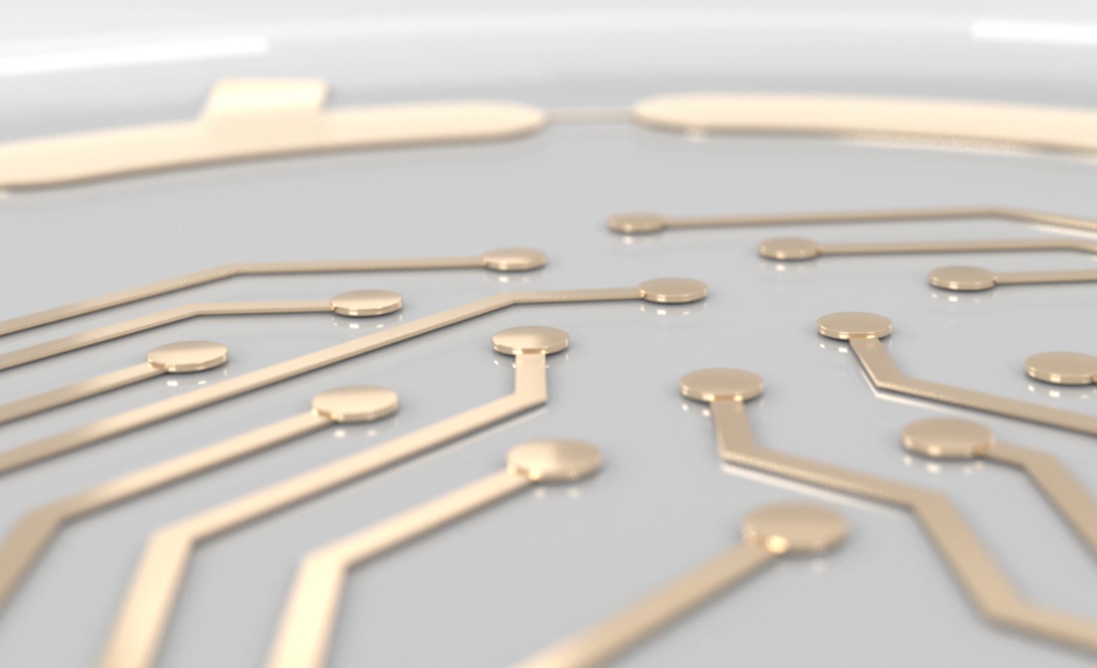
Denser electrode layouts increase the opportunities to sample activity across unevenly distributed, clustered, or sparse cultures. Multiple electrodes may detect the same source, providing information for localization and waveform analysis, when acquisition and signal quality support it.
More stable network-level sampling
Maccione et al. compared high-density hippocampal recordings with subsets of the same area and found that denser sampling could improve the stability of firing and burst estimates and reveal activity sequences. Their results do not establish a universal minimum electrode density for every assay.1
Heterogeneity and putative single units
Population averages can obscure where activity occurs and which groups participate. With appropriate signal quality, spike sorting, and quality control, dense waveforms can support putative unit identification. One electrode is not synonymous with one neuron.
What higher electrode density does not guarantee
More sites do not automatically mean more reliable data
Measurement quality depends on electrode properties, noise, cell-electrode coupling, sampling rate, and analysis. More active electrodes do not compensate for inconsistent plating or replace assay validation.
Total electrodes are not simultaneous recording channels
The selectable sites on a CMOS array may greatly outnumber the channels acquired at once. Sequential mappings cannot by themselves establish truly simultaneous relationships across every sampled location. Compare sites, simultaneous channels per well, and wells recorded in parallel.5
Spatial observations do not replace biological replicates
Additional channels improve observations of a culture but do not increase its independent sample count. Plan donor, differentiation, culture, and replicate numbers separately from electrode density.
Higher planar density is not full 3D tissue coverage
For organoids, electrode geometry and tissue contact matter as much as density. Planar arrays sample the tissue interface rather than the entire organoid volume; different electrode interfaces may be needed for intact 3D preparations. Axion offers specialized organoid MEA technologies for Synexus, including 3DMap, SpheroGuide, and SpheroHD.
3DMap uses flexible electrodes that bend to conform to an organoid, increasing contact across its surface while preserving its three-dimensional shape.
What the literature demonstrates about using both MEA and HD-MEA
Different recording approaches can contribute complementary information to the same biological investigation.
Sundberg and colleagues used patch clamp, MaxOne HD-MEA, and Axion multiwell MEA to investigate human iPSC-derived dopaminergic neurons carrying a 16p11.2 deletion.3
Patch clamp characterized cellular excitability and synaptic events. HD-MEA characterized developing network activity, synchronization, and bursting. Axion multiwell MEA confirmed the network phenotype and supported comparisons involving genotype and treatment.
Rhosin treatment reduced abnormal activity in the conventional MEA experiments. Because treatment occurred during network development, the finding supports modulation of a developing phenotype rather than necessarily acute reversal of an established one.
This study illustrates how different electrophysiological methods can contribute to understanding the same biological model. Detailed spatial recordings can help characterize a phenotype. Targeted measurements can investigate its mechanism. Multiwell assays can expand comparisons across treatments, doses, and biological replicates.
This makes flexibility a practical system-selection consideration.
Synexus supports conventional MEA and HD-MEA within one platform, allowing researchers to select an appropriate recording approach at different stages of an investigation while retaining complementary methods, such as patch clamp, when greater mechanistic detail is required.
How should MEA and HD-MEA measurements be interpreted?
| Measurement | What it can help interpret |
| Firing rate | Overall detected neuronal activity; an observed change does not identify its mechanism alone. |
| Burst frequency/duration | Repeated firing patterns and how they change with maturation or treatment. |
| Network bursts and synchrony | Coordinated timing across a recorded neuronal population and synaptic connectivity. |
| Spatial activity maps | Where signals are detected within the array's actual sampled geometry. |
| Putative single-unit waveforms | Neuronal source discrimination when validated spike sorting and quality control support it. |
| Propagation patterns | Spatiotemporal changes only across locations recorded simultaneously with suitable sampling. |
A change in firing or bursting is not diagnostic of a specific ion channel, synapse, or cell type. Use appropriate controls and complementary methods when mechanistic interpretation is required.
What affects the reliability of MEA measurements?
Reliable recordings require consistent cell preparation and maturation, stable electrode contact, appropriate environmental conditions, adequate signal-to-noise ratio, prespecified detection thresholds, appropriate negative and positive controls, and independent biological replication. Compare workflows using the endpoints and acquisition settings planned for the study.
How to choose a multielectrode array system
Choose a system around your biological question, experimental scale, and complete workflow. Electrode density matters, but so do culture compatibility, simultaneous acquisition, analysis, and the flexibility to pursue new questions.
Define your measurements and experimental capabilities
Conventional MEA and HD-MEA both measure firing, bursting, and network coordination. HD-MEA adds denser spatial sampling for investigating localized activity and heterogeneity. Consider stimulation, microfluidics, custom MEAs, environmental control—including hypoxia alongside recording capabilities.
Complementary measurements can also strengthen interpretation. Synexus combines MEA, HD-MEA, and high-content electrical imaging, allowing researchers to investigate whether functional changes accompany changes in cellular coverage, attachment, or organization.
Match recording coverage to your model
Evaluate electrode density, sensing footprint, culture placement, and tissue contact together. Synexus HD-MEA provides a 4.8 × 4.5 mm sensing footprint, compared with 2 × 2 mm or 0.8 × 0.8 mm for conventional MEA configurations. Cells beyond the array may participate in the network without being directly sampled.
For organoids, electrode geometry is particularly important. Synexus supports specialized organoid MEAs, including 3DMap, whose flexible electrodes conform to the organoid for broader surface contact.
Verify what records simultaneously
Distinguish total electrodes, simultaneous recording electrodes per well, and simultaneous wells. An array with 20,000 electrodes may record only 1,000 at once; channels may also be shared across wells or wells recorded sequentially.
Synexus Pro records across up to 96 wells simultaneously. HD-MEA provides more than 130,000 selectable electrodes and up to 576 simultaneous recording electrodes per well, maintaining that capacity across all 96 wells. Confirm specifications for your intended configuration and mode.
Plan replication—and demonstrate the full workflow
Count donors, independent differentiations, conditions, doses, and replicates. More electrodes improve sampling within a culture; they do not replace independent biological replication.
Request a demonstration at your intended experimental scale. A single-well demonstration cannot establish practical multiwell performance. Assess acquisition time, data volume, analysis time, manual intervention, and endpoint reproducibility across a representative experiment. Synexus is designed around multiwell acquisition and integrated analysis, helping researchers retain experimental scale when adding spatial detail.
Assess culture guidance and scientific support
Review coating, plating, maintenance, recording conditions, and longitudinal handling. Look for protocols and published methods relevant to your model and plate format.
Axion’s culture protocols, application notes, extensive publication library, and scientific support team help researchers identify procedures, optimize assays, and troubleshoot culture or recording challenges.
Compare study costs and future flexibility
Include consumables, software, training, computing, storage, and staff time when estimating study costs.
Synexus lets researchers switch between more affordable conventional MEA plates and HD-MEA plates as experiments require, without a consumable subscription. Configurable software modules and accessories support current needs while allowing capabilities to expand later. Evaluate both the resources needed to complete today’s study and the flexibility to pursue tomorrow’s questions.
Why consider Synexus for conventional MEA and HD-MEA?
Synexus offers the best of both worlds, bringing established conventional MEA workflows and CMOS-based HD-MEA into a configurable multiwell platform. Researchers can match spatial detail to the endpoint, while simultaneously recording across up to 96 wells with Synexus Pro configuration supports controls, treatments, and biological replicates. With appropriate configurations, Synexus with Pixel HD electrical imaging and stimulation or specialized culture options can add complementary experimental capabilities.
Match the MEA measurement to the biological question
Conventional MEA and HD-MEA measure extracellular activity using related principles but offer different spatial sampling options. The right choice depends on the biological endpoint, model geometry, replicate design, and actual recording and analysis workflow.
Synexus combines conventional MEA and HD-MEA with optional complementary electrical imaging, giving laboratories ways to use established network assays and extend to denser spatial analysis when it adds scientific value.
>> Explore Synexus configurations or request a demonstration matched to your model and endpoints.
>> Explore Synexus configurations: [INSERT APPROVED PRODUCT URL]
>> Request an application-specific Synexus demonstration
Related MEA resources
Learn more about MEA technology, applications, protocols, and experimental workflows through these Axion BioSystems resources.
Multielectrode Array Technology
Learn how MEA technology works and how extracellular recordings provide functional measurements from living cells.
Neural MEA Technology
Explore how MEA measures neuronal activity, network behavior, maturation, and treatment responses.
Neural Activity Applications
Explore MEA applications in neural characterization, disease modeling, neurotoxicity, and neural circuit research.
Organoid MEA Technology
Explore electrode interfaces and plate configurations designed for functional recording from three-dimensional biological models.
MEA Culture Protocols
Access culture preparation and experimental protocols for neural, cardiac, and organoid MEA studies.
MEA Application Notes
Review practical assay examples, experimental considerations, and supporting application data.
Scientific Publications and Resources
Explore peer-reviewed studies and technical materials involving Axion BioSystems' electrophysiology platforms.
References and further reading
- Maccione A et al. Experimental investigation on spontaneously active hippocampal cultures recorded by means of high-density MEAs: analysis of the spatial resolution effects. Frontiers in Neuroengineering. 2010;3:4. DOI: 10.3389/fneng.2010.00004.
- Obien MEJ et al. Revealing neuronal function through microelectrode array recordings. Frontiers in Neuroscience. 2015;8:423. DOI: 10.3389/fnins.2014.00423.
- Sundberg M et al. 16p11.2 deletion is associated with hyperactivation of human iPSC-derived dopaminergic neuron networks and is rescued by RHOA inhibition in vitro. Nature Communications. 2021;12:2897. DOI: 10.1038/s41467-021-23113-z.
- Lv S et al. Using Human-Induced Pluripotent Stem Cell Derived Neurons on Microelectrode Arrays to Model Neurological Disease: A Review. Advanced Science. 2023;10:2301828. DOI: 10.1002/advs.202301828.
- Schröter M et al. Advances in large-scale electrophysiology with high-density microelectrode arrays. Lab on a Chip. 2025;25:4844–4885. DOI: 10.1039/d5lc00058k.
- Roberts R et al. Detection of seizurogenic compounds in human and rat neurons in vitro: A multi-laboratory, multi-model assessment. NeuroToxicology. 2026. https://www.axionbiosystems.com/resources/publication/detection-seizurogenic-compounds-human-and-rat-neurons-vitro-multi-laboratory
- Clements M et al. Establishing a functional standard for hiPSC-neural models used to screen for seizure liability with the multielectrode array (MEA) assay. ISSCR poster, July 2026. https://www.axionbiosystems.com/resources/poster/establishing-functional-standard-hipsc-neural-models-used-screen-seizure-liability
- Axion BioSystems. Maestro MEA Platform Powers First Stem Cell Cardiac Safety Assay in FDA ISTAND Program. February 2026. https://www.axionbiosystems.com/resources/news/maestro-mea-platform-powers-first-stem-cell-cardiac-safety-assay-fda-istand-program
- Axion BioSystems. Neural Characterization and Development; longitudinal neural maturation example, data courtesy STEMCELL Technologies (Mak et al., SfN 2016). https://www.axionbiosystems.com/applications/neural-activity/neural-characterization-and-development
Frequently asked questions about MEA and HD-MEA systems
Not necessarily. Conventional MEA provides established network-level measurements and efficient multiwell studies when endpoints are validated. HD-MEA adds denser spatial sampling for questions about localized activity, spatial heterogeneity, and potentially individual units. Choose according to the endpoint, reproducibility, and analysis required.
Manufacturers include Axion BioSystems, MaxWell Biosystems, and 3Brain. Axion's Synexus supports conventional MEA, HD-MEA, and complementary electrical imaging; other systems have various single-well and multiwell configurations but only support HD-MEA. Compare current product specifications and validated workflows.
Yes. Conventional MEA electrodes can detect extracellular action potentials from nearby neurons, although spatial coverage and separating overlapping sources may be limited. HD-MEA can provide additional waveforms for validated localization or spike-sorting analysis.
Not necessarily. Total selectable sites may exceed simultaneously acquired channels. Verify per-well channel counts, sequential versus parallel acquisition, and wells recorded at the same time.
Yes. Multiwell HD-MEA configurations exist, including the Synexus Pro 96-well configuration described here. Always confirm whether wells are actually acquired simultaneously for the planned plate and mode.
Yes. Only Axion BioSystems' Synexus platform supports both conventional MEA and HD-MEA in appropriate configurations, allowing researchers to select established network-level readouts or denser spatial sampling for different experiments.
HD-MEA describes densely spaced sensing sites; CMOS-MEA describes an implementation using complementary metal-oxide-semiconductor electronics. Many high-density arrays are CMOS-based, but the two terms describe different features.


