Authors: Alia Arslanova, Samantha Wong, Sonia Franciosi, Glen F. Tibbits, Maksymilian Prondzynski, and Shubhayan Sanatani
Journal of Arrythmia, 24 September 2026
Longitudinal MEA recordings reveal changes in beat rate and repolarization with relatively preserved conduction in human atrial and ventricular cardiomyocytes exposed to ADHD medications.
The cardiac effects of medications used to treat attention-deficit/hyperactivity disorder (ADHD) remain an important consideration, particularly for patients with underlying heart conditions. Researchers used human iPSC-derived atrial and ventricular cardiomyocytes to investigate responses to methylphenidate, dextroamphetamine, and atomoxetine at concentrations selected to reflect therapeutic plasma exposure. Caffeine and the β-adrenergic agonist isoproterenol were also evaluated to characterize model responsiveness.
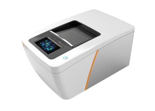
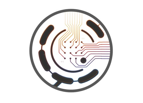
Using Axion MEA, the team recorded extracellular field potentials from spontaneously beating cultures before treatment and at intervals over 48 hours. The AxIS Cardiac Analysis Tool quantified beat rate, corrected field potential duration, and conduction velocity. Repeated measurements from the same wells allowed the researchers to compare acute and prolonged responses in the two chamber-specific cell types.
The ADHD medications produced an early increase in beat rate followed by a decline over time and prolonged corrected field potential duration, most prominently with atomoxetine. Conduction velocity remained largely preserved in both atrial and ventricular cultures. These findings indicate effects on automaticity and repolarization with relatively little change in conduction under the tested conditions. The study supports the use of human cardiomyocyte MEA assays to characterize distinct aspects of cardiac drug response and guide further investigation of potential arrhythmic mechanisms.
Related Products