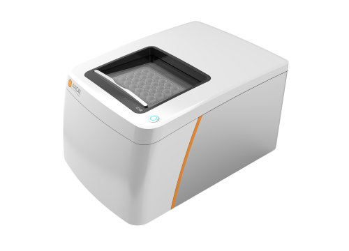
Accurately recapitulating human cardiac activity in the lab is essential for disease research, therapeutic discovery, and safety testing—but traditional animal models often fall short. However, new approach methodologies like stem-cell derived cardiac organoids offer a more human-relevant platform for investigating pathological mechanisms and accelerating preclinical testing. Pairing 3D cardiac organoids with sensitive functional analysis provided by next-generation Maestro MEA offers high-fidelity replication of disease features and dependable assessments of drug responses in vitro.
Learn how Maestro MEA can support your cardiac organoid research with these selected publications:
- Generation of human iPSCs derived heart organoids structurally and functionally similar to heart
- Modeling acute myocardial infarction and cardiac fibrosis using human induced pluripotent stem cell-derived multi-cellular heart organoids
- Evaluation of the cardiotoxicity of Echinochrome A using human induced pluripotent stem cell-derived cardiac organoids
- Frameshift variants in C10orf71 cause dilated cardiomyopathy in human, mouse, and organoid models
Generation of human iPSCs derived heart organoids structurally and functionally similar to heart
Lee S-G, Kim Y-J, et al. Biomaterials. (2022)
Increased demand for physiologically relevant in vitro models has driven the development of organoids and other complex models to study diseases and screen therapeutics. In this study, researchers present and validate a self-organized heart organoid model from human induced pluripotent stem cells (hiPSCs).
Highlights:
>> hiPSC-derived heart organoids display physiologically relevant structures, such as atrium- and ventricle-like areas.
>> Electrophysiological analysis of hiPSC-derived heart organoids using Maestro MEA indicate functional maturation throughout organoid development.
>> Organoids respond as expected to pharmacological treatment with common ion channel blockers, suggesting utility for cardiotoxicology and drug discovery applications.
Modeling acute myocardial infarction and cardiac fibrosis using human induced pluripotent stem cell-derived multi-cellular heart organoids
Song M, Choi DB, et al. Cell Death & Disease. (2024)
The complexity of the adult heart makes it challenging to recapitulate cardiac diseases and injuries, such as myocardial infarction. Here, the authors present a novel human in vitro heart model using self-organizing heart organoids (HOs) containing multiple cardiac cell types. These HOs successfully mimic key pathological features of acute myocardial infarction and ischemia-reperfusion injury, indicating a promising alternative to animal studies for investigating heart diseases and for drug screening.
Highlights:
>> hiPSC-derived heart organoids contain multiple cardiac cell types, including cardiomyocytes, fibroblasts, and endothelial cells.
>> An ischemia-reperfusion model using cobalt chloride provides stable hypoxia induction allowing for more accurate modeling of acute myocardial infarction and reperfusion.
>> Organoids exposed to ischemia-reperfusion conditions exhibit tachycardia, prolonged field potential duration, and decreased spike amplitude and conduction velocity, indicative of significant functional deficits detected by the Maestro.
Evaluation of the cardiotoxicity of Echinochrome A using human induced pluripotent stem cell-derived cardiac organoids
Lee S-J, Kim E, et al. Ecotoxicology and Environmental Safety. (2025)
Despite its therapeutic potential for cardiovascular disease, the cardiac safety of Echinochrome A (EchA) requires further investigation. To address this gap, the authors employed human iPSC-derived cardiac organoids (hCOs) to assess whether EchA causes heart damage or irregular heart rhythms.
Highlights:
>> hiPSC-derived cardiac organoids express key ion channels and markers indicative of functional maturity.
>> The Maestro MEA platform was used to evaluate hCO function as an indicator of cardiotoxicity, finding low cardiotoxic potential with little change in beat period and field potential duration.
>> In addition to supporting the safety and therapeutic use of EchA, this study highlights the translational relevance of cardiac organoids for toxicity assessment.
Frameshift variants in C10orf71 cause dilated cardiomyopathy in human, mouse, and organoid models
Li Y, Ma K, et al. The Journal of Clinical Investigation. (2024)
Dilated cardiomyopathy (DCM) has widely been associated with various genetic mutations. Researchers identified a candidate causal gene, C10orf71, and used hiPSC-derived cardiomyocytes and organoids to assess the effects of C10orf71 mutation on cardiomyocyte electrophysiology and DCM.
Highlights:
>> C10orf71 frameshift mutations are associated with DCM, and C10orf71 is specifically expressed in cardiomyocytes.
>> Contractility analysis of hiPSC-derived heart organoids using Maestro MEA indicates impaired contractile function – evident via decreased amplitude and increased excitation-contraction delay.
>> Contractile protein activation via OM treatment rescues contractile dysfunction in vivo, suggesting therapeutic potential for DCM treatment.


