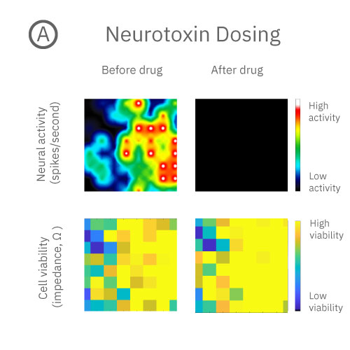
Know what happened to your neurons
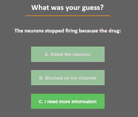
By measuring neural activity alone, you can't tell whether the drug killed the neurons or blocked an ion channel. With the new MEA Viability Module you can stop guessing. Measure both viability and activity in one assay.
In this example, when the drug was added, neural activity was silenced but the neurons are still alive.
How do we know?
With the new MEA Viability Module you can measure both viability and activity in one assay. Get more information from every plate, whether you're modeling development, disease progression, or screening new treatments.
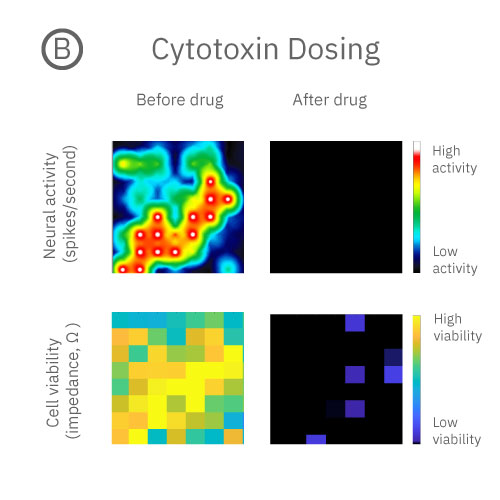
Distinguish functional neurotoxins from cytotoxins in one assay
Functional neurotoxins impact neural activity; cytotoxins impact cell health; both can stop neural activity. With the MEA Viability module, you can distinguish between functional neurotoxins and cytotoxins by measuring both neural network activity and viability in one simple assay in your MEA plate.
Drug dosing can impact activity and viability
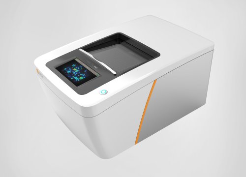
Neural cell coverage and functional activity of hiPSC-derived neurons were tracked using the Maestro Pro multiwell MEA system.
At the start of the assay, both wells showed robust neural activity, network bursting, and cell viability.
Well A was dosed with a functional neurotoxin that silenced neural activity, but did not impact cell viability. Well B was dosed with a cytotoxin which killed them.
Without measures of cell viability, it is difficult to distinguish cytotoxic effects from functional effects. With the new MEA Viability software module, you can see the whole picture, tracking cell viability with Axion's advanced impedance-based technology.
Confirm cell coverage in your MEA experiment
Put away your microscope.
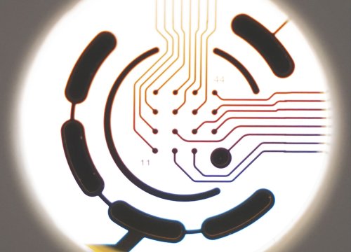
Where are my cells? How’s my cell coverage? How does the structure of culture relate to its functional activity? The MEA Viability Module uses impedance-based technology to quantify viable cell coverage. Coverage in each well is visualized in the Viability Map, with yellow representing denser coverage and blue lower coverage.
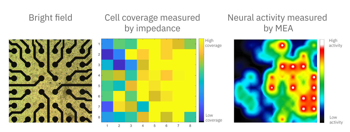
Now you can measure cell coverage and activity in the same assay.
Frequently asked questions about measuring cell viability
Can I record both cell viability and electrical network activity from the same well?
Can cellular viability be measured in high throughput?
Is MEA Viability an endpoint assay?
How does MEA Viability compare to other techniques?
MEA Viability is a non-destructive, dynamic assay. So, there is no need to guess which timepoints to measure, take samples from each well, or transfer the cells to a different system.