Cardiomyocyte Pacing
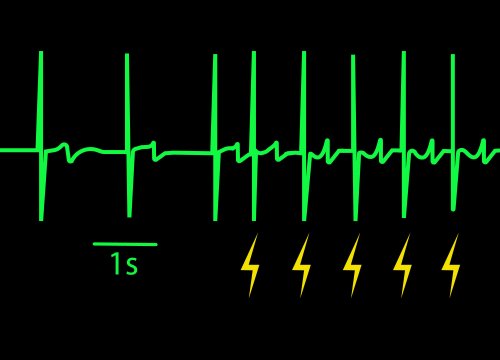
Control cardiomyocyte beat rate with optical or electrical stimulation to increase the physiological relevance of the assay
Learn More→
Pixel HD is the next generation, multimodal, high-density CMOS sensor architecture designed to capture electrical signals from living cells with exceptional spatial detail. Within Synexus, Pixel HD enables HD-MEA and high-content electrical imaging while establishing a scalable foundation for additional electrode-based measurements.
Available exclusively on Synexus.
Live-cell research often separates function, structure, spatial detail, and time across different instruments and experiments. Pixel HD brings these dimensions closer together, helping researchers understand not only whether biology changes, but where the change begins, how it develops, and how it affects the living system.
Pixel HD supports multiple ways of measuring living biology through one high-density electrode interface.
 Cellular-scale spatial analysis — Examine local structural measurements across the entire sensing surface with over 138K electrodes with 12.5 µm spacing in each well of a 96-well plate.
High-definition electrophysiology — Map activity at single-cell scale with up to 576 simultaneous recording electrodes per well across all 96 wells.
Time-lapse phenotyping — Follow live-cell structure, behavior, and response without labels.
Multimodal live-cell analysis — 60+ functional and 20+ structural measurement modes connect function, spatial context, and change over time.
Expandable sensing foundation — Support future electrode-based measurement capabilities.

"We are only beginning to explore what Pixel HD can reveal. I'm excited to see how researchers use its unique capabilities to uncover new insights into cell behavior, function, and response." - Stacie Chvatal, VP Product Management, Axion BioSystems
Pixel HD uses a high-density CMOS electrode array to record signals produced by living cells and measure how cells interact with applied electric fields. These two principles enable high-definition electrophysiology and high-content electrical imaging through the same sensor architecture.
Recording in vitro electrophysiological (MEA) activity
Electrically active cells generate extracellular voltage changes that are detected by nearby electrodes. Dense spatial sampling reveals where activity begins, how it propagates, and how local activity contributes to the behavior of the wider network.
Single-cell morphology with electric fields
For electrical imaging, Pixel HD applies low-amplitude electric fields across the sensing surface. Cells perturb these fields as current encounters cell membranes, cell-cell junctions, and the interface between the cells and electrodes.
Varying the electrical contrast
During each scan, Pixel HD changes the geometry and frequency of the field to produce more than 20 complementary impedance measurement modes. Field geometry changes the direction and path of current, while frequency changes how the field interacts with different parts of the cell-electrode interface.
These combinations provide complementary sensitivity to properties such as:
Turning electrodes into image pixels
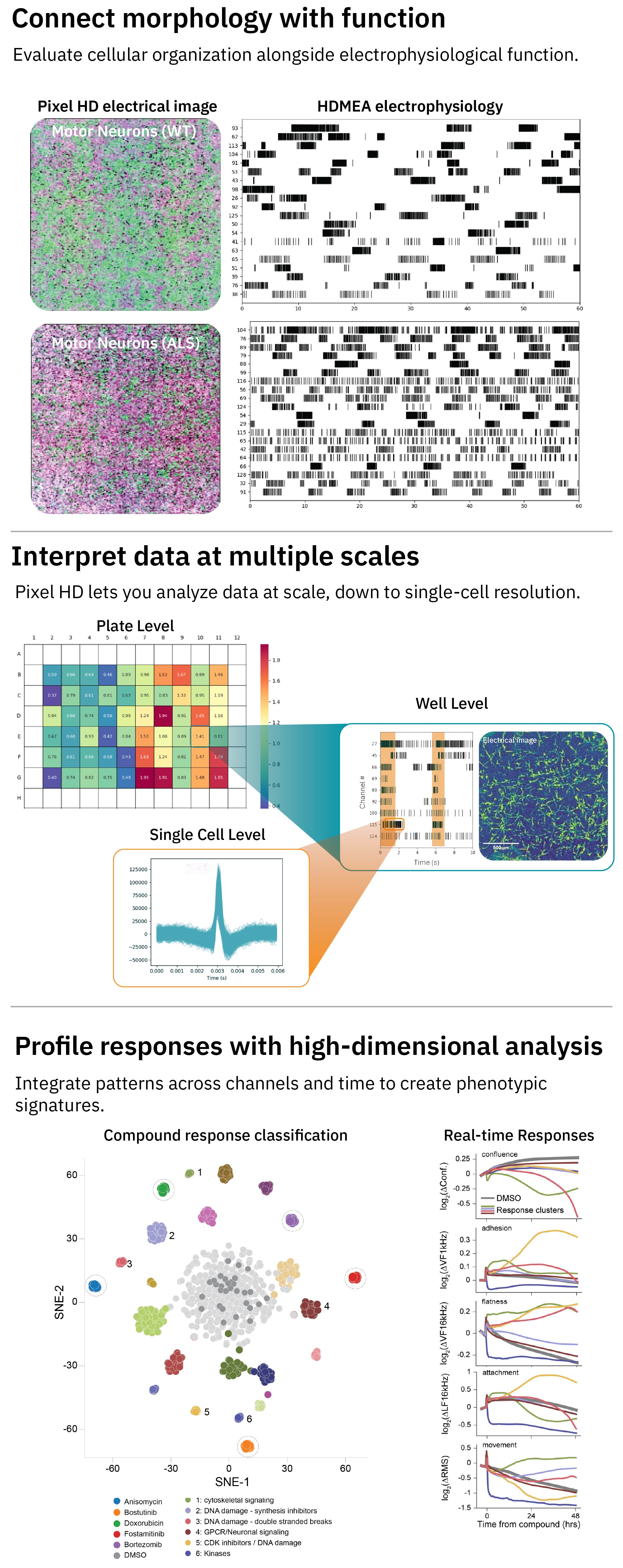
Each electrode occupies a known position and produces a local measurement value. Mapping those values across the sensing surface creates a spatial electrical image in which pixel intensity represents the local electrical response of the cells.
Preserving local variation
Electrode-level measurements can be retained individually or summarized across a well. This allows researchers to examine population averages, spatial patterns, and the distribution of phenotypes within the same culture.
Following change over time
Because the measurement is noninvasive and label free, Pixel HD can repeatedly scan the same culture. Successive measurements create time-lapse views of development, treatment response, toxicity, recovery, and shifting population dynamics.
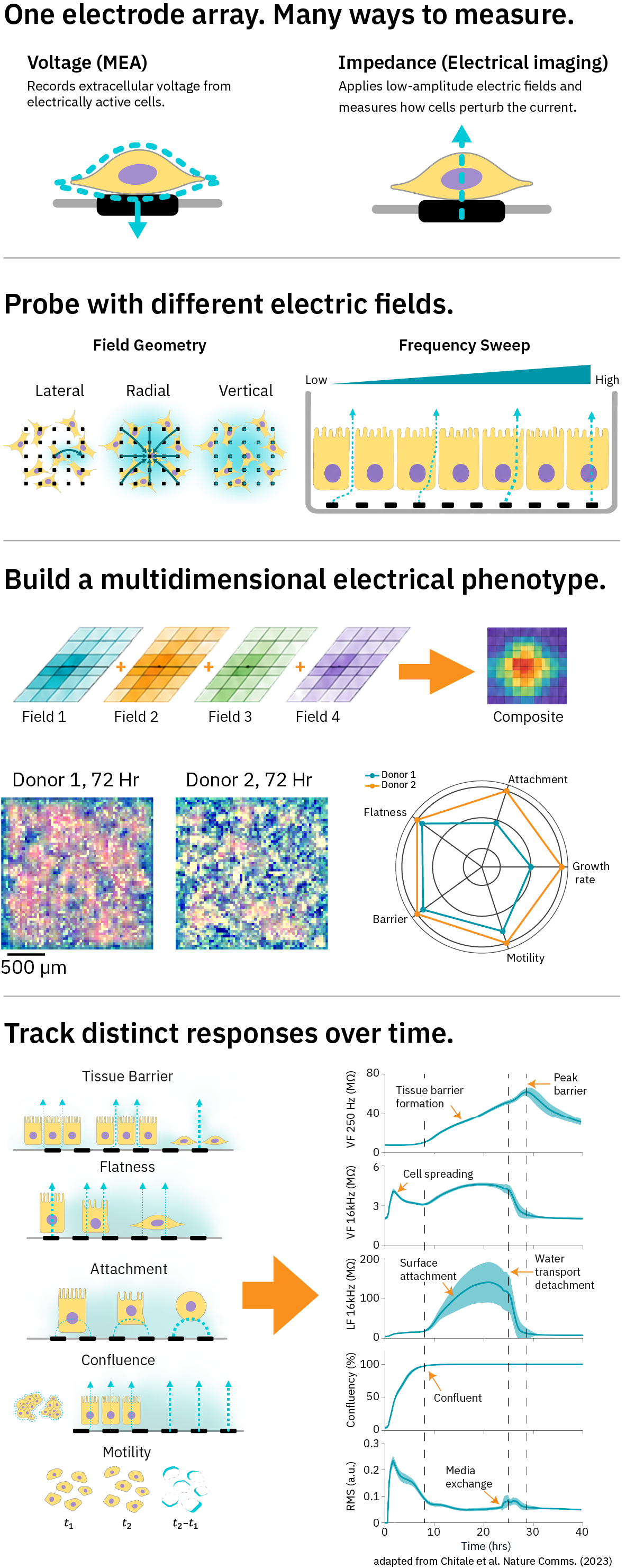
For high-content imaging users, the closest analogy is a multiplexed image stack. Each impedance measurement mode functions as an electrical contrast channel, emphasizing different aspects of cellular structure or behavior.
Examine individual channels
A single channel can make a property such as coverage, attachment, barrier integrity, flatness, or motility more prominent. Each channel has a dominant sensitivity, but it should not be interpreted as a one-to-one molecular marker.
Create composite electrical images
Selected channels can be assigned different pseudo-colors and layered into a multicolored composite. Like a multiplexed optical image, the composite brings several dimensions of the phenotype into one spatial view and makes regional differences easier to recognize.
Unlike fluorescence imaging, these colors do not represent stains, proteins, or fluorophores. They encode different electrical interactions between the cells and sensing surface.
Interpret the data at multiple scales
Resolve heterogeneous populations
Two cultures can produce a similar well average while containing very different local phenotypes. Examining the distribution of electrode values can reveal distinct populations, intermediate states, and changes in population composition that would otherwise be averaged together.
When reference phenotypes are available, the distribution of a mixed culture can be compared with those references to estimate how strongly each population contributes to the overall response.
Build high-dimensional phenotypic profiles
Patterns across channels, distributions, and time can create electrical signatures of cell state and treatment response. These profiles can distinguish phenotypes and reveal different biological trajectories—even when cultures eventually reach a similar endpoint.
Connect phenotype with function
On Synexus, electrical images can be evaluated alongside Pixel HD electrophysiology. This connects changes in cellular organization, morphology, and population behavior with neural or cardiac function.
Pixel HD electrical images are not photographs of the cells. Biological interpretation is strongest when patterns are evaluated across multiple channels, spatial locations, time points, and matched experimental controls.
Different measurement methods answer different biological questions. Pixel HD is designed to add spatially resolved, longitudinal electrical information while complementing established approaches.
Classic MEA provides proven, scalable measurement of functional activity across established assays. Pixel HD adds dense spatial sampling, HD-MEA, and electrical imaging. On Synexus, researchers can access both approaches within one platform.
HD-MEA provides detailed spatial electrophysiology but is often limited to a small number of samples. Pixel HD brings high-density measurement into a scalable multiwell architecture and extends the sensor beyond electrophysiology to electrical imaging.
Endpoint assays provide detailed molecular or biochemical information at selected moments. Pixel HD follows the same living culture over time, revealing kinetics, transient responses, and recovery that may occur between endpoints.