Join Axion's SVP of Marketing, Mike Clements, as he discusses organoid production, analysis, and neural organoid workflow using the Omni cell imaging system. This webinar was originally presented at WORD+2024 - World Organoid Research Day 2024 in Cambridge, UK and is now available for on-demand viewing.
Transcript of webinar:
Thanks for joining my webinar today. I’m going to discuss: Standardizing organoid production and analysis for functional neural disease modeling.
My name in Mike Clements, and I’m a Senior Vice President at Axion BioSystems. I’m a neuroscientist by training with, experience manufacturing stem cell derived models in industry.
Organoids are a powerful new cell model, but there are often 2 major challenges:
- They can be difficult to produce consistently.
- They can be difficult to study in a non-destructive manner. You’ve spent months making your cell model, so the last thing you want to do is to fix and stain it to study it.
At Axion BioSystems, our goal is to help you capture the complex biology of your cells, noninvasively, in real time.
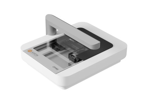
This is our Omni live-cell imager. A simple to use, whole vessel imager, that can sit on a shelf in your incubator. It works like a flatbed scanner, imaging the entire multi-well plate or flask. The major applications for the system are for optimizing cell line development, testing different media formulations, monitoring cell growth, etc.
Why image the entire culture surface? Well when you do you get a more accurate understanding of your cell culture. Here you can see the differences in cell coverage if you on focus on just a small sample area versus the whole well.
And in 2023 we launch a new 12-plate version of the Omni system, called the Omni Pro 12. It has a robotic arm to shuttle the plates down onto the imaging surface and then back into the plate hotel. The experimental scheduler automates this process, reducing the amount of time you need to spend monitoring the progress of your cell cultures.
Now I’m going to quickly cover how the Omni imaging system is being used to help standardize and make the production of stem cell lines and organoids more consistent.
Cell line development can be challenging. For example, confirming that you have dispensed a single cell in a well. Then tracking the speed of proliferation of this colony over time. As you can see, the full vessel scan of the Omni makes this effortless. We visit many labs where this process is still performed manually on a bench top microscope. Prolonged periods out of the incubator is suboptimal for stem cells. It’s also probably not the most effective use of your time.
Here you can see an Omni full plate scan of a 6-well plate. Just like Google Earth, but for your cell culture, you can zoom down to reveal all the colonies in your well. Sliding the scroll bar shows you how these colonies grow over time. And the software will calculate how many colonies are in each well for you, so no more manual counting.
The Omni can detect all the colonies in a well, count them, and track them over time.
The quality of your stem cell culture going into your differentiation will have a major impact on the quality of the resulting cells. Monitoring stem cell culture proliferation can be very time consuming and subjective. Are your cultures growing as fast as previous cultures, is the culture at 50% or 65% confluence. Will I need to passage the cells on Sunday or can I do this on Monday. The new iPSC module on the Omni takes all this guess work off the table, helping you to improve the consistency of your iPSC culture.
The Organoid Module on the Omni can help standardize the embryoid body, formation and expansion process. This can be a critical step in the organoid production process. Here you can see how the software can help you track critical features of embryoid body development including diameter, roundness, and the number in the well.
So, I’ve discussed how Axion is providing tools to help you make more consistent stem cells and organoids. Now I’m going to cover how we are giving researchers deeper insights into their functionality of their neural organoids without disrupting them, using the Maestro MEA system.
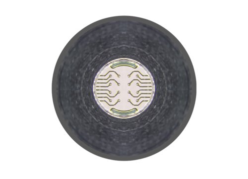
How does Maestro MEA work? Here we’re looking at the culture surface of a multi-well plate. It has a grid of 16 recording electrodes embedded in the culture surface. When neurons are cultured over the recording electrode, the neural action potentials can be detected as changes in the field potential at adjacent the electrode.
And because there is a grid of recording electrodes at the bottom of the well we can detect both spatial and temporal information from the neural network growing in the well. On the right of the screen is a raster plot with electrode #1 at the top and #16 at the bottom. When a neural spike is detected a check mark is added to the raster plot giving us a visual display of the neural network activity. This network activity can change as the network develops, in response to genetic edits, and application of test compounds.
And as there is an electrode array in each well of the multi-well plate it means that you can use this assay to test multiple test conditions with time matched positive and negative controls as you would for any other assay. With the major applications being studying cell development, cell characterization, disease modeling and screening for therapies.
Why is there so much interest in recording from neural organoids on Maestro MEA? This seminal paper from Cleber Trujilio in Alysson Moutri’s Lab demonstrated the complex neural network activity that develops in neural organoids. After 4 months in culture, nested oscillations were detectable in the neural burst firing, which mimicked the types of activity seen in the EEG from the developing human brain.
What they also showed was that this same complex neural activity was not present when the same cells were used to make neurospheres, or 2D monolayers. It was only present in the self assembling neural organoids.
Now over 40 publications have already been published on Maestro MEA using neural organoids covering a wide range of neurological disorders including Alzheimer’s, ALS, Autism, Epilepsy, Glioblastoma, and Parkinson’s Disease.
To make neural organoid recordings on Maestro even easier, we have recently launched our new SpheroGuide MEA plate. The well design funnels the organoid over the recording electrodes in the plate enabling faster and more consistent loading of the plate. We can confirm that the organoid is touching the electrodes using the impedance mode on the Maestro system, and then switch to neural activity mode for the electrical activity recordings.
Here is a video showing how we can confirm, using impedance, that the organoid is touching the electrode array in the well. With the neural module we can then detect the spontaneous electrical activity from the organoid and the complex burst firing patterns. As the Maestro MEA plates are transparent, when combined with our Omni imaging system, we can seamlessly multiplex the imaging and functional data, so you have the most complete understanding of your neural organoid.
Finally, with need for even higher through put, our customers are starting to automate their Maestro MEA assays. Here is an example from Revvity, where they integrated the Maestro Pro system with their plate::works 6.30 scheduler software. This integration enables automated recordings and workflows with liquid handlers and other instruments.
So in summary, at Axion we want to provide you with the tools to help you make more consistent stem cells and organoids, and then give you deeper insights into their functionality without disrupting them.
And as a point to leave you on, organ-on-a-chip models provide another approach for creating more physiologically relevant in vitro cell models. Axion has recently partnered with the microfluidics experts at Nerti to produce the first multiwell microfluidic plate with MEA electrodes integrated into it.
Here we can see neurons seed in one compartment project neurites out and across into the neighboring compartment. The electrical activity in the microfluidic chip can be recorded using the Maestro Pro system.
And that all for today’s webinar. If you have any questions, please feel free to contact us at info at axionbio.com and we’ll get back to you shortly.