Quick summary: Watch the webinar to explore how tumor heterogeneity, immune donor variability, and alternative immune checkpoint pathways are being investigated to better understand immunotherapy response and resistance in hepatocellular carcinoma.
Why do some hepatocellular carcinoma models respond to immune checkpoint inhibition while others remain resistant?
Hepatocellular carcinoma (HCC) is characterized by substantial molecular and immune heterogeneity, which can influence how tumors interact with the immune system and respond to immunotherapy. Understanding this variability requires looking beyond the presence or absence of a single immune checkpoint.
In this webinar, researchers explore the biological factors that may influence response and resistance to immune checkpoint inhibition in HCC. Learn how PD-L1 regulation, tumor molecular subtype, immune donor variability, Aurora kinase A, and alternative immune checkpoints including PVR and Nectin-2 can shape immune-mediated tumor cell killing and inform the investigation of combination strategies.
What will you learn:
- How PD-L1 expression and regulation vary across HCC models
- Why tumor molecular subtype can influence PD-L1 stability and immune-mediated killing
- How immune donor variability affects response to PD-L1 checkpoint inhibition
- How real-time impedance measurements reveal the kinetics of tumor cell killing
- How Aurora kinase A inhibition may influence PD-L1 regulation and immune susceptibility
- Why PVR and Nectin-2/CD112R are being investigated as alternative immune checkpoint pathways
- How tumor-intrinsic and immune factors can inform the study of combination strategies
Why does immunotherapy response vary in hepatocellular carcinoma?
Hepatocellular carcinoma exhibits substantial intertumoral and intratumoral heterogeneity. Tumors classified under the same disease can have different molecular programs and immune environments, potentially leading to different responses to treatment.
Immune checkpoint inhibitors targeting pathways such as PD-1/PD-L1 can restore immune-mediated tumor killing by blocking inhibitory signals that suppress activated immune cells. However, responses remain heterogeneous, suggesting that checkpoint expression alone does not fully explain whether a tumor will respond.
The webinar explores this complexity by examining both sides of the tumor-immune interaction: tumor-intrinsic biology and characteristics of the immune effector cells.
PD-L1 regulation extends beyond gene expression
PD-L1 is often considered in terms of whether it is expressed by a tumor, but the webinar demonstrates why its biology can be more complex.
In the HCC samples examined by the researchers, PD-L1 mRNA levels did not differ substantially between tumor and paired non-tumoral tissue. At the protein level, however, a different pattern emerged: PD-L1 protein was increased in a subset of HCC samples.
The researchers also identified multiple PD-L1 molecular species representing different maturation and glycosylation states. The mature, highly glycosylated form was associated with the plasma membrane and was increased in HCC tissue in their analysis.
These findings illustrate why measuring total PD-L1 alone may not capture functionally relevant differences between tumor models.
How does HCC molecular subtype affect PD-L1?
The researchers next investigated whether PD-L1 heterogeneity was associated with HCC molecular subtype.
In the cell models presented, the S1 subtype exhibited higher PD-L1 expression, including greater membrane-associated PD-L1, while the S2 model showed lower expression. The experiments also suggested differences in how the two subtypes maintained PD-L1 over time: the S1 model more effectively stabilized mature PD-L1, whereas the S2 model exhibited faster turnover and greater reliance on continued protein synthesis.
These differences were accompanied by distinct responses when the tumor cells were co-cultured with activated immune cells.
Using real-time impedance measurements on the Maestro Z, the researchers observed that the S1 model with higher, more stable PD-L1 was more susceptible to immune-mediated killing, while the S2 model with lower PD-L1 showed greater resistance and slower killing kinetics.
The result highlights an important theme of the webinar: PD-L1 expression needs to be interpreted within the broader tumor and immune context.
Immune donor variability influences checkpoint inhibitor response
Tumor biology was only part of the observed variability.
To model the immune component, peripheral blood mononuclear cells (PBMCs) from healthy donors were activated and co-cultured with HCC cells. The researchers then evaluated responses to the PD-L1-targeting antibodies atezolizumab and durvalumab.
Continuous impedance measurements enabled tumor cell viability and response kinetics to be followed throughout the experiment. Despite using the same tumor cells, experimental conditions, and checkpoint inhibitor, responses differed according to the PBMC donor.
Some donors showed enhanced tumor killing following PD-L1 blockade, while others showed little additional benefit. When experiments were repeated using independent blood samples from the same donors, the response patterns remained reproducible.
Rather than treating donor-to-donor variability simply as experimental noise, these findings suggest that the characteristics of the immune compartment can be an important experimental variable when investigating checkpoint inhibitor response.
How does inflammatory context affect PD-L1 blockade?
The researchers also examined whether changing the inflammatory environment could alter the response to checkpoint inhibition.
Interferon gamma increased membrane PD-L1 expression in the HCC model used in the webinar. Although increasing an inhibitory checkpoint might initially appear counterproductive, interferon gamma also reflects and promotes an inflammatory, immune-active environment.
After interferon gamma pretreatment, a donor that had previously shown a limited response to checkpoint blockade became substantially more responsive.
This finding further illustrates why PD-L1 upregulation cannot necessarily be interpreted in isolation as increased immune suppression. Its biological significance can depend on the surrounding inflammatory and immune context.
Exploring Aurora kinase A as a tumor-intrinsic target
If resistance is influenced partly by tumor-intrinsic biology, targeting a tumor vulnerability could potentially alter its susceptibility to immune-mediated killing.
The researchers investigated Aurora kinase A, a mitotic kinase they observed to be increased and activated in HCC. Pharmacological inhibition disrupted mitotic progression, promoted polyploidy, and reduced HCC cell viability in the models presented.
Their experiments also suggested a relationship between Aurora kinase A and PD-L1 regulation. Aurora kinase A inhibition reduced the mature membrane-associated form of PD-L1, although the researchers explicitly caution that their data do not establish direct ubiquitination or stabilization of PD-L1 by Aurora kinase A.
Aurora kinase A inhibition also increased HCC cell susceptibility to activated PBMC-mediated cytotoxicity.
When combined with PD-L1 blockade, however, the outcome again depended on immune context. Additional tumor cell elimination was observed in a donor for whom checkpoint inhibition alone had limited effects, whereas relatively little additional benefit was seen in a donor already responsive to checkpoint blockade.
The researchers therefore do not interpret these findings as universal synergy. Instead, the results support continued investigation into which tumor and immune contexts may benefit from a combination strategy.
Beyond PD-1/PD-L1: Alternative immune checkpoint pathways in HCC
Limited response to PD-1/PD-L1 blockade may also involve compensatory immune escape mechanisms.
The second portion of the webinar investigates members of the Nectin family, particularly PVR (CD155) and Nectin-2 (CD112), as potential alternative immune checkpoint pathways in HCC.
PVR can interact with inhibitory immune receptors including TIGIT and CD96 as well as activating receptors such as DNAM-1. Nectin-2 shares some interacting partners and also binds the inhibitory receptor CD112R. The researchers investigated whether targeting these pathways could enhance immune-mediated HCC cell killing.
Targeting PVR to enhance immune-mediated HCC cell killing
The researchers observed PVR expression across the HCC cell lines they examined, including models in which PD-L1 was absent. This provided a rationale for investigating PVR as an alternative checkpoint target, particularly in PD-L1-negative models.
In co-culture experiments, blocking PVR enhanced NK cell-mediated killing of HCC cells and restored expression of the activating receptor DNAM-1 on NK cells.
The researchers then moved to activated PBMCs from healthy donors. Using real-time impedance measurements with the Maestro Z platform to continuously monitor tumor cell proliferation, they observed suppression of tumor cell growth following PVR targeting across multiple experiments.
These results support further investigation of the PVR pathway as an immune checkpoint mechanism in HCC.
Investigating Nectin-2 and CD112R
Nectin-2 was also highly prevalent in the HCC tissue samples examined in the webinar. Expression was significantly higher in neoplastic tissue than matched distal liver tissue, and higher tumor-associated Nectin-2 expression was associated with reduced early disease-free survival in the cohort presented.
The researchers subsequently investigated whether targeting CD112R, an inhibitory receptor for Nectin-2, could enhance immune-mediated HCC cell killing.
In PBMC co-culture experiments, the effect of CD112R targeting was relatively modest and highly donor dependent. Experiments using NK92 cells provided additional evidence that targeting the pathway could enhance immune-mediated tumor cell killing, but the response was not consistent across all immune effector populations.
These findings reinforce a broader theme running throughout the webinar: the effectiveness of immune checkpoint targeting can depend strongly on the biological and immune context in which it is evaluated.
Real-time monitoring of tumor-immune interactions
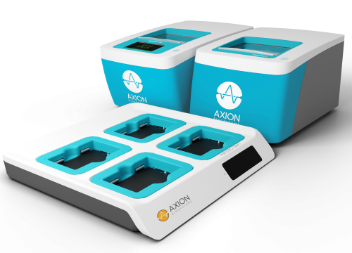
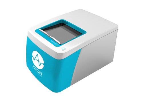
Several experiments presented in the webinar use the Maestro Z impedance platform to follow HCC cell viability and proliferation during co-culture with immune cells.
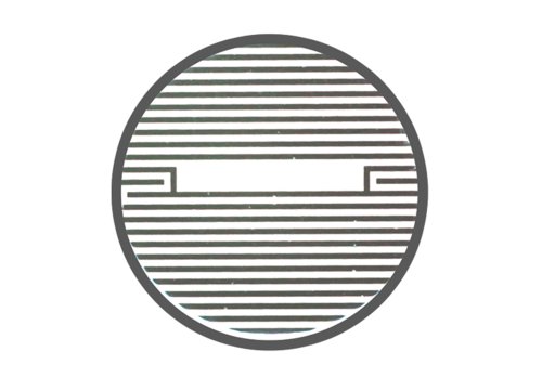
Unlike endpoint measurements that provide a single observation after a predetermined period, continuous impedance measurements allow researchers to observe when immune-mediated killing begins, how the response develops, and how treatment effects differ over time.
This kinetic information was particularly useful when comparing responses among PBMC donors and evaluating checkpoint-targeting strategies whose effects changed throughout the experiment.
Explore real-time assays for immuno-oncology research to learn more about monitoring immune cell-mediated cancer killing in vitro.
Toward more context-specific models of immunotherapy response
The research presented in this webinar demonstrates that response to immune checkpoint targeting in HCC cannot be understood from a single tumor marker or pathway alone.
PD-L1 maturation and stability differed between HCC models. Immune-mediated killing and response to PD-L1 blockade varied among PBMC donors. Aurora kinase A inhibition altered tumor cell biology and immune susceptibility, while PVR and Nectin-2/CD112R provided additional immune checkpoint pathways with distinct response patterns.
Together, these findings support the use of experimental models that incorporate both tumor-intrinsic and immune determinants when investigating immunotherapy response and potential combination strategies.
As the presenters conclude, the research question becomes more specific than simply asking whether an immune checkpoint inhibitor works: which tumor, with which immune context, and under which combination of conditions is most likely to respond?
About the presenters:
Paola Kučan Brlić, PhD,
Assistant Professor and Research Scientist at Center for Proteomics, Faculty of Medicine, University of Rijeka, Croatia
Paola Kučan Brlić, PhD, is an Assistant Professor at the Center for Proteomics, Faculty of Medicine, University of Rijeka, and a researcher in translational immunology, with a focus on tumor–immune interactions and immune checkpoint biology. She obtained her PhD in Biomedicine and Health from the University of Rijeka, where her research centered on the role of the poliovirus receptor (PVR/CD155) in viral immune evasion.
Her current work integrates basic and translational approaches to investigate mechanisms of immune escape in cancer. Her research focuses on immune checkpoint molecules, particularly the nectin family axis, exploring their role in shaping tumor immunogenicity and response to immunotherapy. In particular, her work includes the development of novel therapeutic antibodies targeting the nectin-family axis, with the aim of establishing new immune checkpoint–based treatment strategies.
Dr. Kučan Brlić has contributed to multiple peer-reviewed publications in the fields of immunology and oncology and has been actively involved in international research collaborations. The work presented in this webinar was generated during her postdoctoral research at Fondazione Italiana Fegato (Trieste, Italy), where she investigated nectin family immune checkpoint inhibitors in the context of hepatocellular carcinoma (HCC).
Her research aims to identify predictive biomarkers and develop novel therapeutic strategies, including antibody-based approaches, to improve immunotherapy outcomes.
Luca Grisetti, PhD,
Postdoctoral Researcher at National Institute of Gastroenterology-IRCCS "Saverio de Bellis", Castellana Grotte, Italy
Dr. Luca Grisetti is a Postdoctoral Researcher at IRCCS "S. de Bellis," a national oncology and gastroenterology research hospital, in collaboration with the Italian Liver Foundation. He earned his PhD in Molecular Biomedicine from the Università degli Studi di Trieste in 2024, with a thesis on Aurora kinase A in HCC development and PD-L1 regulation.
His research focuses on the molecular mechanisms driving HCC, with expertise in Aurora kinase A signaling, PD-L1 biology, and immune checkpoint response — advancing precision immuno-oncology for liver cancer. He is the author of 8 peer-reviewed publications in journals including Frontiers in Immunology, Cell Proliferation, and Cancers, and has presented at leading international conferences including EASL, ILCA, and AISF.
His work has earned several awards, including the ILCA Young Investigator Shark Tank Award (1st Prize, 2023) and the AISF Young Investigator Best Poster Award (1st Prize, 2023), and recognition among the Top 5 PhD Theses at the University of Trieste (2023–2024). He currently serves as a Y-ILCA Committee Member (2024–2027) and peer reviewer for JHEP Reports, The International Journal of Biological Markers, and Cancer Biomarkers.