One electrode array. More ways to measure.
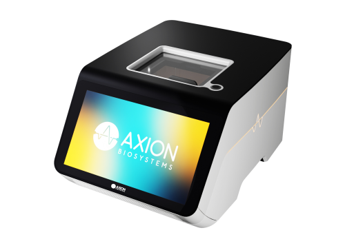
Available exclusively on Axion BioSystems’ Synexus™ platform, Pixel HD is the next-generation CMOS technology for high-definition electrophysiology and high-content electrical imaging. Pixel HD is a semiconductor-to-live-cell interface that captures multimodal, real-time measurements from the same culture.
High-dimensional, high-resolution data delivers the spatial detail to investigate local differences and the ability to follow responses over time, offering new insights into the structure and function of your cells.

High-definition electrophysiology
Measure cellular electrical activity with single-cell resolution.
High-content electrical imaging
Follow cellular phenotype without labels or disruptive endpoints.
Different measurements. More biological detail.
Pixel HD™ varies the frequency and geometry of applied electric fields to generate more than 20 complementary impedance measurement modes. Each mode provides a different sensitivity to cellular properties, adding information about morphology, coverage, attachment, barrier integrity, and other aspects of cell behavior.
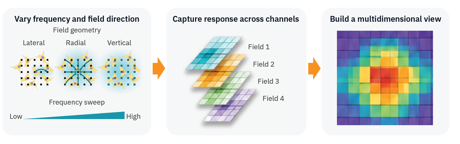
Selected channels can be layered into composite electrical images. Patterns across channels, spatial locations, and time build high-dimensional phenotypic profiles, revealing differences that a whole-well average can hide.
See more in every response.
Electrical imaging connects cellular phenotype with function and monitors change over time.
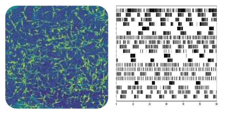
Neural activity & morphology
Explore how cellular organization and local responses relate to electrophysiological function as you track spikes and network behavior together with electrical images.
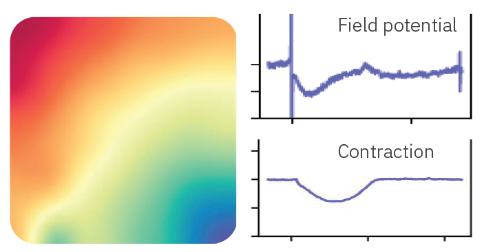
Cardiac function & safety
Connect high-definition conduction, contractility, and field potentials to structural changes in cardiac cultures.
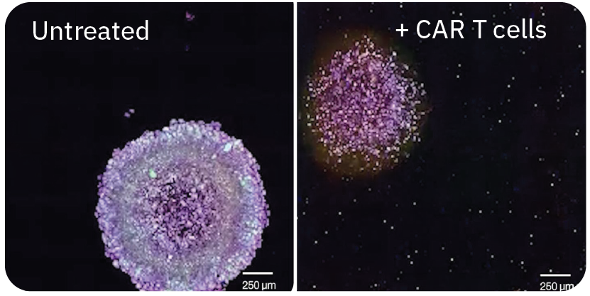
Immune-cell killing & 3D cultures
Follow target-cell responses in 2D and 3D cultures. Electrical imaging adds spatial context to response kinetics, helping researchers examine where cellular changes occur and how they develop.
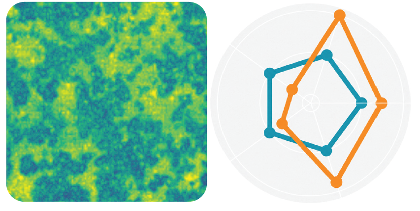
Phenotypic profiling & cocultures
Combine complementary measurements into rich phenotypic profiles, helping distinguish cell populations within cocultures and reveal differences beyond visual appearance. Label-free measurements let researchers follow these populations and their responses over time.
And more.
Explore barrier integrity, attachment, motility, and other label-free impedance assays in high definition.
Related Products