Key Findings
>> The Maestro Z measures antibody-drug conjugate mediated cancer cell killing in a real-time, label-free manner.
>> HER2-targeted ADCs kill HER2high SKOV3 cells more effectively than HER2low A549 cells.
>> The cytotoxic capacity of different ADCs can be compared using the Maestro Z.
Abstract
Antibody-drug conjugates (ADCs), particularly those targeted toward the tumor antigen HER2, have emerged as a popular and effective treatment for cancer patients. Early in vitro assessment of the cytotoxic capabilities of ADCs can inform research and development efforts, as well as give insight into future clinical outcomes. Here, we use impedance measurements with the Maestro Z to assay HER2-targeted ADC killing in a real-time, label-free manner.
Introduction
Numerous immuno-oncology studies have focused on developing cancer therapies that specifically target tumor cells while without damaging healthy tissue. Comprised of a tumor-targeting antibody bonded to a cytotoxic payload, antibody-drug conjugates (ADCs) have emerged as promising and effective therapies, particularly for breast cancers expressing human epidermal growth factor 2 (HER2)1.
Approximately twenty percent of breast cancer patients overexpress HER2, making it an attractive target for targeted therapies2. Trastuzumab is a widely characterized antibody that targets HER2 and, therefore, has been incorporated into ADC products including Enhertu® (trastuzumab-deruxtecan) and Kadcyla® (trastuzumab-emtansine)3.
To assess the effectiveness of ADC therapies early in development, accurate and reliable in vitro methods to assay ADC cytotoxicity are needed. Traditional cell viability assays rely on dyes or fluorescent labels that can be laborious to use. Further, these assays typically provide endpoint readouts and do not allow for continuous monitoring of cell killing. Therefore, information about the dynamics of ADC cytotoxicity may be lost using these traditional methods.
The Maestro Z uses electrodes embedded in the bottom of the well to measure cellular impedance and can detect cell detachment during cell death in a label-free manner. The Maestro Z measures this process in real time, providing a measurement every minute and revealing the precise kinetics of ADC killing. Here, we utilize the capabilities of the Maestro Z to measure the cytotoxic effects of HER2-targeted ADCs on two different cancer cell lines: SKOV3 and A549. Our results reveal the differing kinetics of two clinically available ADCs, Enhertu and Kadcyla. We also show the increased cytotoxic effects of HER2-targeted ADCs against the HER2 overexpressing SKOV3s versus the HER2low A549s. Taken together, this study demonstrates the ease and effectiveness with which the Maestro Z can assay in vitro ADC cancer cell killing.
Materials and Methods
Cells and reagents
SKOV3 (Cat. HTB-77) and A549 (Cat. CCL-185) were obtained from ATCC (Manassas, VA). SKOV3 media was composed of McCoy's 5A Medium Modified (ATCC, Cat. 30-2007), 10% FBS (Gibco, Cat. 16000044), and 1 % penicillin/streptomycin (Gibco, Cat. 15140122). A549 media was composed of F12-K base media (Gibco, Cat. 1127022), 10% FBS (Gibco, Cat. 16000044), and 1% penicillin/streptomycin (Gibco, Cat. 15140122).
Enhertu (trastuzumab-deruxtecan, Cat. D4001), Kadcyla (trastuzumab-emtansine, Cat. D4003), and trastuzumab (Cat. A2007) were purchased from Selleckchem. IgG control antibody was purchased from Invitrogen (Cat. 31154).
Maestro Z assay platform
The Maestro Z platform (Axion Biosystems) uses impedance measurements (ohms, Ω) to quantify the presence of cells on electrodes embedded in the bottom of the wells of CytoView-Z plates (Axion Biosystems). Cellular impedance is a well-established technique for measuring cell attachment, spreading, proliferation, coupling, membrane integrity (cell death), and subtle changes in cell conformation. Detection is noninvasive and label-free, so it can quantify dynamic cellular responses over minutes, hours, and days. The Maestro Z's built-in environmental chamber finely controls temperature and CO2, ensuring a consistent, optimal experimental environment.
Cell plating
CytoView-Z 96 plates (Axion BioSystems) were coated with 100 µL of fibronectin solution (1 µg/mL) per well and incubated at 37°C and 5% CO2 for at least one hour. After incubation, excess surface coating was aspirated from each well. Then 100 µL of SKOV3 or A549 medium was added per well and docked on the Maestro Z platform as a media reference.
Cancer cell lines were thawed and cultured in their respective media according to the supplier recommendations, passaging as needed. Cells were lifted from flasks and dissociated via trypsinization. The cell suspension was then transferred to a 15 mL conical tube and centrifuged at 1,000 rpm for five minutes. The supernatant was aspirated, and cell density and viability were determined using a hemocytometer. SKOV3 and A549 cells were plated at 5,000 cells per well in a 96-well CytoView-Z plate. Cells were added at 100 µL per well, for a total well volume of 200 µL.
The plate was allowed to rest at room temperature for one hour prior to docking on the Maestro Z, which incubated at 37 °C and 5% CO2. Integrated humidity reservoirs on the CytoView-Z 96 were filled with sterile water to maintain humidity.
ADC Dosing
ADCs were serially diluted from stock solutions in the appropriate cell media. At 24 hours post cell seeding, Enhertu or Kadcyla were added at 10x the final concentration to treatment groups. Trastuzumab and IgG were added at the same concentration as controls. A media only addition was also included as a “No Treatment Control” (NTC).
Calculation of cytolysis
%Cytolysis was used to quantify cell death and was calculated using the following equation:
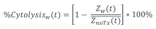
where where ZnoTx(t) is the mean across the No Treatment control wells' time series.
Generating dose response curves in AxIS Z
The dose response analysis in AxIS Z was used to perform a best fit regression using the Hill equation, a 4-parameter logistic model:
where a and b are the minimum and maximum asymptotes, respectively. The dose at half-maximal response (c) is often referred to as the EC50 or IC50. Finally, the Hill slope (d) gives the slope at the steepest part of the curve.
Results
Enhertu kills SKOV3 cells in a dose-dependent manner
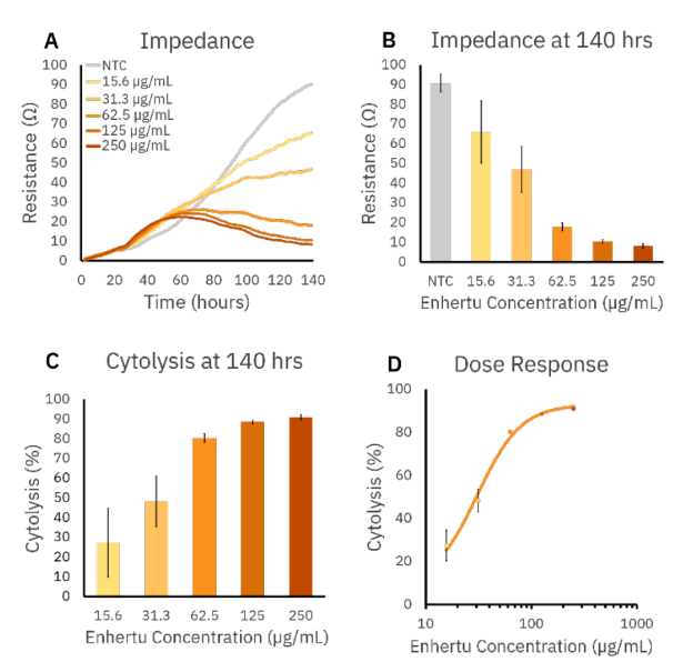
Trastuzumab-based ADCs for treating cancers that overexpress HER2 have shown success in the clinic1. Therefore, we dosed SKOV3 cells, an ovarian cancer line that overexpresses HER2, with increasing concentrations of Enhertu, a clinically available ADC targeting HER2. We plated SKOV3s and allowed them to adhere and proliferate for 24 hours. We then dosed with increasing concentrations of Enhertu and monitored the real-time killing of SKOV3 over 116 hours (140 hours total for the experiment) on the Maestro Z.
Impedance measurements from the Maestro Z showed dose-dependent killing of SKOV3 cells by Enhertu (Figure 1A and 1B), with the largest dose (250 µg/mL) leading to the lowest SKOV3 resistance. The same was true when cytolysis was calculated using the no treatment control (NTC). Increasing concentrations of Enhertu led to increased cytolysis of SKOV3 cells. We used these cytolysis values to produce a dose-response curve and found the EC50 value for Enhertu to be 31 µg/mL. Together, these results demonstrate that ADC in vitro killing can be measured in a real-time, label-free manner on the Maestro Z.
Kadcyla kills SKOV3 more effectively than A549 cells
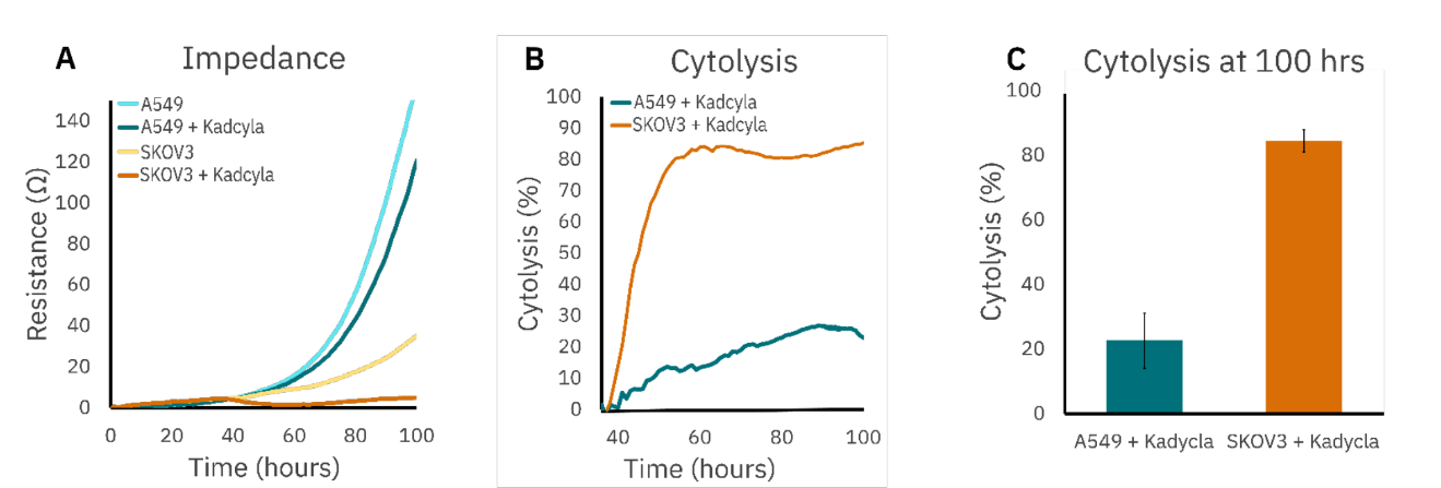
When developing ADCs, it is important to consider the antigen that the ADC's antibody is targeting and its expression levels in both tumorous and normal tissue. If the antigen is overexpressed in tumors compared to healthy tissue, then ADCs can be designed to effectively target the tumor cells without harming healthy tissue. However, lower expression of this marker in cancer cells may lead to less effective tumor killing or unwanted off-target killing4.
To illustrate this point, we dosed SKOV3s, a HER2 overexpressing cell line and A549s, a cell line with low levels of HER2, with another HER2- targeted ADC, Kadcyla. Following dosing with 3 µg/mL Kadcyla at 24 hrs, SKOV3 cells were killed more quickly and completely than A549 cells as shown by resistance measurements taken on the Maestro Z (Figure 2A). SKOV3 cytolysis levels reached 85.3% at 100 hrs post-plating (76 hrs post dose), while A549 cytolysis only reached 22.9% (Figure 2B and 2C). These results highlight the sensitivity of the Maestro Z to discern differences in ADC cytotoxicity based on target cell antigen expression.
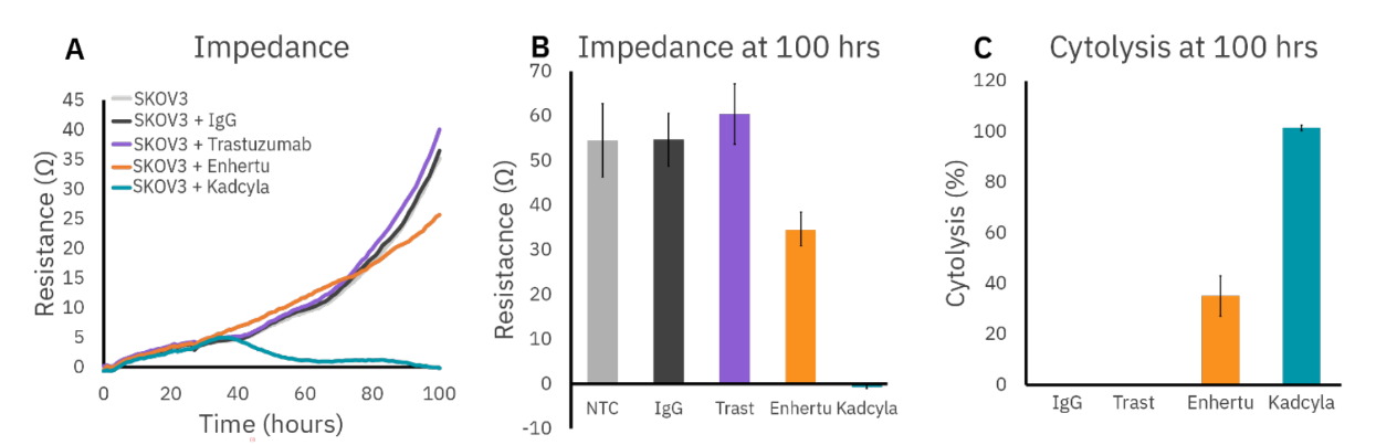
Kadcyla kills SKOV3 cells faster than Enhertu
Comparing different ADC formulations (different cytotoxic payloads, e.g.) is an important part of the therapy development process. Accurate in vitro methods for evaluating ADC effectiveness may help researchers make decisions earlier in the development pipeline that save time and resources further downstream (animal studies, clinical trials, etc.). To illustrate this, we compared the killing of SKOV3 cells by Kadcyla and Enhertu head-to-head on the Maestro Z (Figure 3). Real-time impedance measurements show that Kadcyla kills SKOV3 cells faster, and, at 100 hours, had caused more complete cell death compared to Enhertu (Figure 3A-C). Both ADCs had greater cytotoxicity than control IgG antibodies or trastuzumab alone, which showed no in vitro cytotoxicity of SKOV3 cells5.
Conclusion
Advances in immuno-oncology research have led to several novel and effective cancer treatments, including antibody-drug conjugates. To accelerate development of new ADC therapies, accurate, reliable in vitro assays to assess their cytotoxicity are needed. In this study, we demonstrated the Maestro Z's ability to measure ADC cytotoxicity in a real-time, label-free manner. We were able to generate dose-response curves and calculated EC50 values for ADC killing, as well as resolve differences in cytotoxicity against cell types with different levels of antigen (HER2) expression. Finally, we measured the killing dynamics of two ADCs head-to-head to reveal which was more effective against a given cell type. The methods presented here can be implemented in ADC development pipelines to rapidly identify effective ADC therapies and inform future downstream studies.
References
1. Rassy, E., Rached, L. & Pistilli, B. Antibody drug conjugates targeting HER2: Clinical development in metastatic breast cancer. Breast 66, 217–226 (2022).
2. Wolff, A. C. et al. Human Epidermal Growth Factor Receptor 2 Testing in Breast Cancer: American Society of Clinical Oncology/College of American Pathologists Clinical Practice Guideline Focused Update. J Clin Oncol 36, 2105–2122 (2018).
3. Lewis, G. D. et al. The HER2-directed antibody-drug conjugate DHES0815A in advanced and/or metastatic breast cancer: preclinical characterization and phase 1 trial results. Nat Commun 15, 466 (2024).
4. López de Sá, A. et al. Considerations for the design of antibody drug conjugates (ADCs) for clinical development: lessons learned. Journal of Hematology & Oncology 16, 118 (2023).
5. Yu, L. et al. Eradication of Growth of HER2-Positive Ovarian Cancer With Trastuzumab-DM1, an Antibody-Cytotoxic Drug Conjugate in Mouse Xenograft Model. International Journal of Gynecological Cancer 24, 1158–1164 (2014).
Authors
Benjamin Streeter, Senior Scientist
Denise Sullivan, Senior Applications Manager
Daniel Millard, VP of R&D - Bioelectronics
Axion BioSystems, Atlanta, GA