What is Live-Cell Imaging?
Live-cell imaging is an indispensable method to observe and quantify dynamic biological processes in real time and covers a wide range of applications, including the analysis of cell health, proliferation, cell migration, morphology, and function.
Benefits of Live-Cell Imaging
Live-cell imaging provides the most stress-free environment for your cells – the incubator. Instead of bringing samples to an instrument for measuring, we bring the instrument to your cells. This allows the capture of sensitive, time-dependent events that can otherwise be missed using traditional, endpoint assays.
With live-cell imaging you can:
- >> Observe dynamic cellular processes over time
- >> Minimize disruption of cellular biology
- >> Reduce hands-on time
- >> Gain new insights with AI-supported analysis
With brightfield and fluorescent capabilities, our live-cell platforms are compatible with a wide variety of assays and applications.
See More, Guess Less
Whole-well brightfield imaging can improve the reliability of your experiments, ensuring your results aren’t skewed by an uneven plating. While generating and analyzing this kind of data is usually time consuming, Axion’s advanced image analysis tools can rapidly get an unbiased image-based analysis of cell behavior and function.
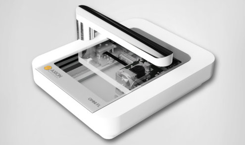
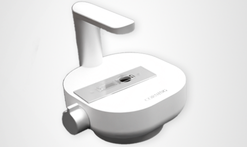
Explore Axion's Imaging Platforms
Take your Maestro system even further with powerful add-ons designed to extend functionality and unlock new experimental possibilities.