Inotropy
Characterize compound-induced effects on cardiomyocyte inotropy and excitation-contraction coupling
Learn More→
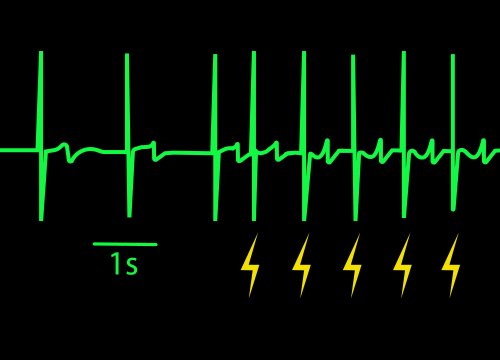
Every beat of the heart is characterized by a contraction of the heart that pumps the blood out to the body. When cardiomyocytes are cultured on top of electrodes, they form a spontaneously beating syncytium. With each beat, the cells contract and relax, changing their shape and coverage over the electrodes. These changes can be measured as a change in impedance, or contractility.
Contractility is often used to characterize the mechanical properties of induced pluripotent stem cell-derived cardiomyocytes and to detect the effects of compounds on cardiac contractile function (e.g., inotropes). Measures such as beat amplitude, beat period, and excitation-contraction delay reveal changes in contractile function due to cardiomyocyte maturation or compound addition.
![]() Noninvasive, impedance-based recording of cardiomyocyte contraction without dyes or labels
Noninvasive, impedance-based recording of cardiomyocyte contraction without dyes or labels
![]() High temporal and spatial resolution to capture single-electrode and well-wide measurements
High temporal and spatial resolution to capture single-electrode and well-wide measurements
![]() Long-term monitoring of cardiomyocyte function, including changes in contraction due to drug response
Long-term monitoring of cardiomyocyte function, including changes in contraction due to drug response
![]() Multi-well, scalable formats suitable for comparative and screening studies
Multi-well, scalable formats suitable for comparative and screening studies
![]() Multiplexing with electrophysiological data to measure excitation-contraction coupling
Multiplexing with electrophysiological data to measure excitation-contraction coupling