Biocompare, 2021
Apoptosis, a form of programmed cell death, plays an important role in removing damaged cells from healthy tissue, and in removing unnecessary cells during development. Apoptosis can occur via an intrinsic or extrinsic pathway, but a malfunction of either can result in diseases such as cancer or neurodegenerative conditions. Because apoptosis is not a single event but a process with many stages, it pays to consider your needs before choosing an apoptosis assay from among the multitude available. This article discusses some common apoptosis assays and provides expert advice on choosing them.
Common apoptosis assays
Each apoptosis assay tests for a particular point in the process, with assay methods—often flow cytometry, western blotting, microplate reader, or agarose gel electrophoresis—that are convenient for the user and best for the cell type under study. Here are some commonly used assays for different stages of apoptosis.
Early-stage assays. Two common assays for early apoptotic events are the mitochondrial membrane potential assay and the cytochrome c assay. Early in apoptosis, pores open in mitochondrial membranes, making them permeable to ions and other molecules. The resulting depolarization of the mitochondrial membranes can be assayed using fluorescent indicator dyes. In addition, when pore formation disrupts mitochondrial membrane potential, cytochrome c is then released from mitochondria into the cytoplasm. The assay quantifies cytochrome c in the cytosolic versus mitochondrial extracts.
Early-to-mid stage assays. Two widely used early-to-mid stage apoptosis assays are for annexin V and caspase. During apoptosis, phosphatidylserine moves from the inner to the outer layer of the cell’s plasma membrane. An indicator of this stage of apoptosis is the presence of annexin V (conjugated to a fluorescent dye), which binds to phosphatidylserine, on the cell surface. Caspase activity is another indicator of apoptosis; because members of the caspase family of proteolytic enzymes vary in their roles in the process (for example, initiator caspases are active earlier than effector caspases), it’s helpful to know which caspases are likely relevant to your inquiry.
Late-stage assays. DNA fragmentation is a late-stage indicator of apoptosis, and is commonly detected by DNA laddering on agarose gels, TUNEL assays, or via DNA binding dyes in flow cytometry.
Considerations when choosing assays
Sometimes scientists choose a particular apoptosis assay simply because it’s well-known and validated, such as annexin V, says Dmitry Kuksin, Head of Reagent Development and Commercialization at Nexcelom Bioscience. But it’s not necessarily the best apoptosis assay for their particular experiment, which might require more than one assay to test for cells in different stages of apoptosis. “When you talk about apoptosis, you want to be very specific about the percentage of cells that are positive in different apoptosis assays,” says Leo Li-Ying Chan, Head of Advanced Technology R&D at Nexcelom Bioscience. “These measurements are complementary and tell different parts of the story, and that’s why scientists are using all of them to study drug effects on cancer cells, for example.”
Nexcelom Bioscience (a PerkinElmer company) offers both instruments and reagents for apoptosis detection. The Cellometer K2 and Cellometer Spectrum platforms can run different types of apoptosis assays in various formats using fluorescence detection, such as annexin V, propidium iodide, and caspase. In addition, the Celigo Image Cytometer, and the automated Cellaca MX high-throughput cell counter—both with brightfield imaging and 4+ fluorescent channels—are geared toward high-throughput plate-based assays.
Promega, which offers tools for apoptosis assays detecting DNA fragmentation, caspase activity, and phosphatidylserine exposure, advises scientists to consider the assay sensitivity they require, and the hands-on time devoted to the assay workflow. “Our homogeneous, plate-based assays are well-suited for drug discovery because of speed, simplicity, and scalability into high-density well formats,” says Andrew Niles, Senior Research Scientist at Promega. “However, our TUNEL and imaging formats require more effort but are often entirely sufficient for exploration of basic science questions where fewer samples are used and at a lower price point.” Further considerations include cell culture type, such as cells in suspension, 2D or 3D cultures, and whether other biomarker assays are required. “We offer reagent solutions tailored to meet these specific requirements with live-cell kinetic methods that offer multiplexing compatibility,” says Niles.
Orthogonal confirmation of apoptosis
Confirming apoptosis using more than one type of assay is often advisable, and sometimes even required for peer-reviewed publication. “Although many parallel methods can provide corroboration for apoptosis, we have found same-well multiplexing to be highly desirable due to efficiencies in the workflow and lower intrinsic experimental variation,” says Niles. For example, Promega’s RealTime-Glo™ Annexin V Apoptosis and Necrosis assay, directly followed by the Caspase-Glo® 3/7 Assay reagent, can be performed within the same well. “This method is possible because of reagent addition sequence and optimization of the detection chemistries,” says Niles. “This combination of assays notably delivers data for two well-established parameters for determining apoptosis.”
Apoptosis plus other parameters
Assay choice may also involve consideration of multiple parameters in addition to apoptosis. “For example, in doing a drug screen, customers are now trying to couple a lot of these assays together,” says Kuksin. “They're looking for functionality, they're looking at viability, growth rate, apoptosis, and cell cycle, trying to get a full picture to understand, for example, what’s my drug’s mode of action, is it initiating an apoptosis cascade, or is it simply arresting the cell cycle?” Multi-drug treatments that mimic the combination of drugs received by a chemotherapy patient can pose a new host of challenges, as some cells positive for an apoptosis marker may be in stasis but still recover, whereas others might continue on down the apoptotic pathway to die. “At this point, they start looking for additional markers, dyes, and reagents in order to get a much bigger and better overall picture of their culture,” notes Kuksin.
For scientists who want to know the time course of the apoptotic response, a kinetic study might reveal answers. “Most cell biologists now recognize that the stimulus’s mode of action and inherent potency dictate the rate of progression through the apoptotic program,” Niles says. Unfortunately, such quantities are often unknown. “Our real-time annexin V assay can definitively establish the kinetics, potency, and magnitude of apoptotic responses by repeated collection of data points throughout the entire exposure.”
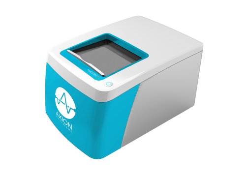
Tracking changes in cell health is also important when examining the timing of events within cells, which can help to identify stages of apoptosis in multiple pathways. “Some [apoptotic] markers are expressed only in certain pathways and, even then, only transiently,” says Steven Fiene, Scientific Liaison at Axion Biosystems. “Knowing the correct markers, drug concentrations, exposure times, and the kinetics of the cellular response are critical, [because if you] take measurements too early or too late, you may not see a response at all.”
Axion Biosystems’ Maestro Z live-cell analyzer continuously tracks changes relevant to apoptosis, such as cell attachment, membrane integrity, and cell death. It uses impedance-based measurements for real-time, continuous label-free monitoring of cell cultures. “Because the Maestro Z assay requires no labels and doesn't interfere with the cell culture in any way, it can be multiplexed with other apoptosis assays, providing kinetic cell health data to help put the mechanistic apoptosis assay results in context,” says Fiene.
There is no “best apoptosis assay” because each individual research situation is unique. However, considering the stage(s) of apoptosis, additional health or kinetic parameters, cell type, and assay format should move you further toward choosing the best apoptosis assay(s) for your research.