Sixty-five million people world-wide suffer from epilepsy. Epilepsy is characterized by seizures, which are the result of excessive nerve activity in the brain. While treatments are available, finding the optimal medication for any patient involves a period of trial-and-error. The need to better understand the underlying causes and develop better treatments for epilepsy are clear. In this webinar, Dr. Mike Boland and Dr. K. Melodi McSweeney (Columbia University) discuss the power of utilizing genetically engineered mice to explore the neuronal networks associated with epilepsy.
Audio Transcript:
Sixty-five million people worldwide suffer from epilepsy. Epilepsy is characterized by seizures which are the result of excessive nerve activity in the brain. This condition is a range of disorders that varies from mild to severe some of which can be life-threatening, while treatments are available finding the optimal medication for any patient involves a period of trial and error during which, both doctor and patient will try to find a solution that improves their quality of life. Unfortunately for about 1/3 of them the prescribed treatment will not work, we need to better understand the underlying causes and develop better treatments for those suffering from epilepsy.
In today's coffee-break webinar Mike Boland and Melody McSweeney discuss how they are harnessing the power of genetic engineering in mice to explore the neuronal networks behind epilepsy. With a better understanding of these brain networks, it could be possible to classify different types of epilepsy, allowing for the most effective medication to be prescribed. For the first time without further ado, I'd like to introduce you to today's researchers Dr. Michael Boland is an assistant professor at Columbia University where he serves as director of the cellular models of disease initiative with the Institute for Genomic Medicine. Melody McSweeney is a PhD candidate currently studying genetics with an emphasis on neurodevelopmental disorders, both she and Dr. Boland have worked extensively with David Goldstein, as all three of them dig deeper into the medical mysteries of neuro diseases.
Thanks for the introduction Melissa as mentioned Melody and I are based at the Institute for genomic medicine at Columbia University. Columbia University recently introduced precision medicine initiatives in five key areas of medicine; undiagnosed childhood diseases, kidney and liver disease, maternal-fetal medicine, ALS, and epilepsy. At the heart of each of these initiatives is the Institute for genomic medicine which was created to serve as the face of these efforts. The IGM, under the direction of human geneticist David Goldstein, uses state-of-the-art whole exome and whole sequencing coupled with advanced bioinformatics to accurately identify disease-causing genetic variants. The IGM is a highly collaborative multidisciplinary Institute comprised of genetic counselors that interface with patients clinicians and IGM leadership, it also consists of a genomic sequencing core facility and a team of bioinformaticians. Additionally, the IGM possesses a disease modeling group that focuses on neurodevelopmental disorders, such as autism intellectual disability, and epilepsy. We use human induced pluripotent stem cells derived from patients as well as human iPS cells and mouse lines generated by genomic editing, to accurately represent the genotype of patients. These models are subjected to a number of technical platforms that are designed to identify and characterize genotype-specific phenotypes, some of these platforms are shown here.
Our disease modeling group collaborates with other academic groups and industry leaders to identify and develop new therapeutics, here we'll focus on using microelectrode arrays to study activity patterns of primary neuronal networks from mouse models of epilepsy. The IGM under the direction of Wayne Frankel has developed 14 mouse models of epilepsy from genes that affect a range of biological processes such as the function of neuronal ion channels or transporters, the endocytosis of synaptic vesicles, synaptic function, signaling through cell surface receptor, and RNA processing. We've developed corresponding human IPS L models for many of them to more accurately study cellular phenotypes we use microelectrode arrays as the primary screening tool to examine neuronal networks from these models for genotype-specific phenotypes and for recurrent phenotypes across models.
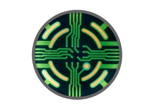
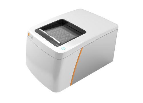
Briefly, MEAs are tissue culture wells embedded with multiple electrodes that measure local field potentials generated from electrically active cells, software then converts that information into an activity profile for each electrode. We use Axion's multiwell MEA Maestro system equipped with a Lumos module for optogenetic manipulation of networks.
MEA has a number of advantages over traditional wholesale patch-clamp electrophysiology, it represents a non-invasive method to record network activity from populations of neurons rather than single cells. It allows for a researcher to monitor network development and establishment, and also to test several experimental conditions in parallel. The medium throughput of the multiwell format allows for small-scale compound screening or testing. The IGM has developed a custom MEA analysis package that's freely available to the research community and can be found at the web address at the bottom of the slide.
As a proof-of-concept, we investigated whether changes in excitability could be identified using the MEA following modulation of neuronal network activity. A number of recent studies have implicated certain micro RNA's in epilepsy, for example, when the expression of micro RNA 128 is knocked down, it results in seizures in mice and death within about three months of age. We used a lentivirus to deliver a micro RNA inhibitor called a sponge RNA which is comprised of tandemly repeated sequences that are partially complementary to the mature micro RNA. We use the sponge RNA to knock down expression of micro RNA 128 in primary mouse cortical neurons, and then investigated changes in network excitability. A schematic of the micro RNA sponge is shown on the left, it is expressed downstream of a GFP reporter gene.
On the right, we see cells expressing the green fluorescent protein on an MEA well. The video is an example of recorded network activity, each box represents one well of a 12-well MEA plate. Each dot is one of the 64 electrodes, in each well the intensity of the colors reflect the activity of the nearby neurons. You can see that the six control wells on the left fire much less frequently than the wells treated with the micron a sponge on the right. A descriptive way to visualize this data is to utilize raster plots which demonstrate activity from each electrode over time. We generated raster plots using our in-house MEA analysis package to show that in the control wells there are well-defined bursts that synchronize across most electrodes and well-defined interest intervals in which there is no activity.
The network's treated with the micro RNA sponge shown on the right fire more frequently and have little and highly variable time between bursts. We further analyze the activity data using our pipeline and identified significantly increased mean firing, rate burst rate, and synchronous network spikes in wells in which micro RNA 128 expression was reduced, we conclude that suppression of micro RNA 128 expression results in network hyperexcitability in vitro. Given this proof-of-concept data, we moved on to test this paradigm by evaluating primary neuronal cultures on MEAs using one of our genetic mouse models.
This spider plot shows a clear fingerprint of genotype-specific activity patterns in one of our epilepsy models. When compared to wild-type non-epileptic cells shown here in black and given the value of one, the mutant network shown here in blue, has a similar number of active electrodes and mean firing rate which are measures of network health and activity. The mutant networks however, have significantly increased number of bursts, decreased percent of spikes and bursts, mean duration of burst, and mean interest interval. This means the epileptic network has many short very rapid bursts with very little time between them.
The mutant network also has increased number of network spikes and percent of spikes in network events, which are features of synchronicity. This illustrates hyperexcitability and hyper synchronicity in the mutant. Raster plots nicely illustrate this aberrant network activity, on the left is a representative activity plot from a wild-type Network compared to a representative mutant network on the right. Both types of networks possess defined bursts and inter burst intervals but, the mutant networks exhibit shorter bursts with very short into burst intervals. Also note that the bursts in the mutant network have a fixed burst duration and rigid periodicity, in comparison to controls. We recognize this phenotype from our experiments involving pharmacological manipulation of neuronal subtypes in networks. In particular, this phenotype is reminiscent of GABAergic inhibition. When neurotransmission in wild-type networks is modulated by GABAA receptor antagonists such as GABA seen we observe significant increases in mean firing rate bursts per minute and percent of spikes inverse, which is reminiscent of our epilepsy mutant networks.
This data provides some insights into the underlying mechanism responsible for the aberrant phenotypes observed in the mutant networks. Ultimately our goal is to use MEAs to identify small molecules or drugs that are capable of correcting or ameliorating mutant phenotypes so they resemble wild-type activity.
We've shown that MEAs represent a viable tool to identify and analyze genotype-specific network phenotypes.
Furthermore, ornithologist disease models based on solid human genetics represent better models for mechanistic studies and therapeutic design. The multiwell MEA platform is amenable to medium throughput drug screening and lead candidate compounds from large screens can be prioritized based on MEA findings, and these findings can be used for phenotype correction or amelioration before being tested in an animal.
We'd be remiss if we didn't acknowledge David Goldstein, Aaron Hinesin and Wayne Frankel, as well as other members of the IGM and funding agencies. Thank you for your attention.
And, that wraps up today's coffee-break webinar, if you have any questions you would like to ask regarding the research presented or if you are interested in presenting your own research with microelectrode array technology, please forward them to coffeebreak@axionbio.com for questions submitted for Dr. Boland and Melody McSweeney, they will be in touch with you shortly.
Thank you for joining me on today's coffee break webinar and we look forward to seeing you again!


