Cell culture protocol for recording from CDI iCell Cardiomyocytes2 on Maestro MEA systems
Cell Culture Protocol
FCDI iCell Human iPSC Cardiomyocytes
Preparing the MEA Plate
- Place a 5 μl droplet of fibronectin over the recording electrode area of each well in the MEA plate. See Figure 1 on page 2 for appropriate drop placement.
Reconstitute fibronectin in sterile PBS at 1 mg/ml. Aliquot and store at -20° C, and dilute to 50 µg/ml the day of use.
Reccomend to add 6-8 mL of sterile water to the on-plate reservoirs to increase humidity.
- Incubate the fibronectin-coated MEA plate in a cell culture incubator at 37°C, 5% CO2 for at least 60 minutes.
Culturing iCell Cardiomyocytes
- Thaw iCell Cardiomyocytes2 according to the iCell Cardiomyocytes2 User’s Guide.
- Remove a sample of the cell suspension and count the cardiomyocytes using a hemocytometer to determine both the viability and total number of viable cells. Transfer the cell suspension to a 15 ml conical tube.
Ensure the cardiomyocytes are evenly suspended before removing an aliquot to count.
- Centrifuge the cell suspension at 180 x g for 5 minutes.
- Aspirate the supernatant, being careful not to disturb the cell pellet.
- Dilute the cell suspension in Plating Medium to 10,000,000 viable cardiomyocytes/ml.
Plating iCell Cardiomyocytes onto the MEA
- Aspirate the fibronectin solution from the MEA.
- Place a 5 µl droplet of iCell Cardiomyocytes2 suspension (approx. 50,000 cells) over the recording electrode area of each well of the MEA that has been aspirated. See Figure 1 on page 2 for appropriate drop placement.
Timing is critical in this step. Cardiomyocyte attachment is compromised if the fibronectin is allowed to dry after aspiration. Therefore, best practice is to aspirate and plate cells one row at a time. If the residual fibronectin has turned white and crystallized, the well should be ignored.
- Incubate the MEA plate with the seeded cardiomyocytes in a cell culture incubator at 37°C, 5% CO2 for 1 hour.
- Gently add 1/2 of the final volume of Maintenance Medium to each well of the MEA. Adding the medium too quickly will dislodge the adhered cardiomyocytes. Recommended well volumes for each plate type are: 6- and 12-well = 1000 µL, 24 = 500 µL, 48-well = 300 µL, 96-well = 200 µL.
Using a pipette, add medium first in a semi-circle along the outer edge of the well. Progressively add medium to either side of the well so it fills evenly towards the center. The goal is to prevent a rush of medium in either direction that might dislodge the cardiomyocytes.
- Repeat step 11 a second time to reach the final recommended volume of Maintenance Medium.
- Incubate in a cell culture incubator at 37°C, 5% CO2 .
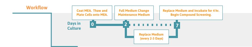
- For optimal cell health, perform a full media change on Day 2 and then exchange 50% of the medium every 2-3 days. Though cardiomyocyte beats may be detectable within 24 hours, optimal Na+ spike and T-wave amplitudes are typically achieved after at least 7 days in culture.
Drop Placement
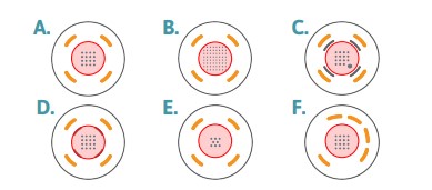
Figure 1: Drop Placement Diagram
The layouts above represent the bottom surfaces of wells in (A) a 48-well MEA, (B) a 6- or 12-well MEA, (C) a 24-well MEA or 48 well E-Stim+ MEA, (D) a 48-well AccuSpot MEA, (E) a 96-well MEA, and (F) a 48-well CytoView MEA. The number of electrodes per well is different across the plate formats, however the drop placement is the same, with the drop (red circle) centered on the recording electrodes and staying within the ground electrodes. On plate types (C) with the addition of the stim-paddle in the lower right corner of the array, it is important to make sure the droplet covers this feature. The droplet may need to be manipulated after placement of the pre-treatment to ensure stim-paddle coverage.
Visualization of Typical Cardiomyocyte Seeding Results
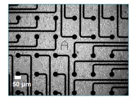
Figure 2: iCell Cardiomocyte Morphology
Human iPSC-Derived Cardiomyocytes2 (50,000) at day 9 in vitro in a 12-well CytoView MEA, 10x magnification. Notice that cardiomyocyte morphology is easily recognizable.
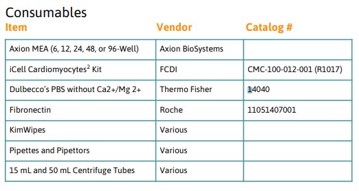
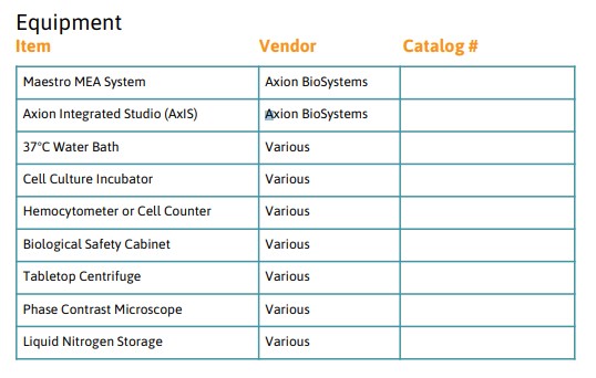
Required Materials