|
2023 |
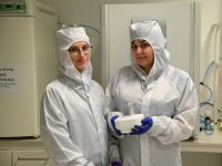
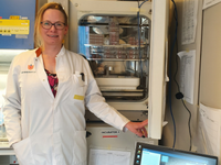
Paulina Trzaskowska, Warsaw University of Technology
Device: Lux 3 FL
Statement: "The scientific project involving the use of the Lux device involves real-time studies on the differentiation of different cell types using bioactive agents. The research will be conducted on two cell types: human mesenchymal stem cells (hMSCs) and human mammary fibroblasts (HMFs). The hMSCs from bone marrow are capable of differentiating in various ways, including cartilage tissue. The project will investigate the effect of TGF-B3 growth factor content in the culture medium and culture time on hMSCs differentiation, including changes in cell morphology and proliferation, assessed using real-time observations. HMFs are primary cells that can differentiate into CAFs (cancer-associated fibroblasts). The project will involve real-time observation of the change in shape, size and migration of these cells."
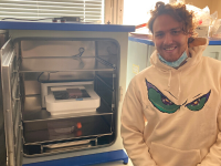
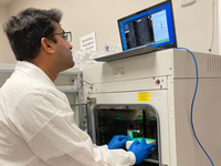
Alex Gray, UCL - London
Device: Omni FL
Statement: "We are using the Omni FL to help dissect multi-drug resistance in cancer. Live-cell imaging allows us to track the growth and behaviour of patient-derived cells; monitor 3D spheroids and their responses against drug panels and analyse the complex cellular mechanisms that cause resistance. The Omni's time-lapse feature tracks the formation and collapse of our 3D cancer models whilst the fluorescent channels are used to follow the movement of substances in and out of the cells as well as tracking cells localisation in co-cultures."
|
2022 |
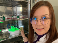
Adriana Memete, University of Oradea
Device: Lux 3 BR
Statement: "In our project, we proposed to study whether anthocyanin-rich fruit extracts have a protective effect on UV irradiated fibroblasts and wound healing potential effect. With the help of Lux3 BR we will be able to visualize in real time the different biological events that can occur in the treated cells. Live-cell imaging under cell culture conditions will allow us to observe and understand the events that take place in the cells, e.g. morphological changes, cell confluence, wound healing rate. Another advantage of using the device is that the images or videos of the cells can be monitored and analyzed from another location, without involving the opening of the incubator. "
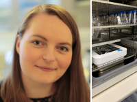
Simone Porsborg, Aalborg University
Device: Omni
Statement: "In Regenerative Medicine at Aalborg University, we investigate stem cells and replacement tissues to harness their regenerative potential and synergies with the ultimate goal of developing novel therapeutic approaches and translating these into novel regenerative therapies. In doing this, we use a plethora of predictive models to mimic the tissue response, and by using the Omni we hope that both the technical and biological quality of these models will be even better."
Judy van Beijnum, AUMC
Device: Omni
Statement: "Very excited to start working with the Omni device! Within the Angiogenesis Laboratory at AUMC, we investigate novel types of immunotherapies to target the tumor vasculature, including vaccination and CAR T cells. With the Omni, we plan to develop assays to monitor angiogenic tube formation and sprouting, as well as CAR T cell activity, Furthermore, we aim to improve cell migration analysis readouts and algorithms."
Tanmay Mathur, BioInSyst Lab
Device: Lux 3 FL
Statement: "The BioInSyst Lab is a leader in the development of organ-on-chip biotechnology and innovating the next generation of preclinical cellular systems of human diseases. The impact of our research is far reaching as organ-chips mimic complex disease states and offer a testbed for drugs."
David Wallace, Oklahoma State University
Device: Lux 3 FL
Statement: "The Lux3 FL allows simultaneous real-time measurements of different biomarkers including oxidative stress and apoptosis, helping to characterize the genesis of pancreatic cancer. With this understanding, we can begin to screen potential therapeutics that will attenuate or block the development of pancreatic cancer."
|
2021 |
Leon Schurgers, Maastricht University
Device: Lux 3 FL
Statement: "We are very excited to receive the Lux3 FL. We are now able to easily visualize important intracellular trafficking of labeled proteins in iPSC-derived cardiomyocytes. By altering signaling pathways, we aim to elucidate the molecular mechanisms of cardiovascular diseases. Using patients-derived iPSC-lines, we are able to differentiate the iPSCs into beating cardiomyocytes with a background of different diseases, with and without a gene-corrected control."
Ary Marsee, Utrecht University
Device: Omni
Statement: "Using the Omni 384-well plate scanner, we could track the morphological changes (branching, growth, condensation, fusion, collapse, etc.) during the self-organization of organoids from single cells and disordered cell clusters over time. The data generated using the Omni will be used to instruct culture parameters and develop a standardized workflow for organoid culture."