What you will learn in this 8 minute webinar:
- >> Dr. Tomasello describes a novel electrophysiological technique for measuring neural activity in live zebrafish larvae.
- >> This multielectrode array (MEA) based assay can be easily adapted by labs with no prior ephys experience.
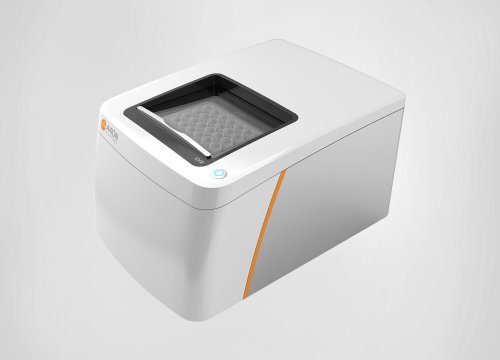
- >> The Maestro Edge system provides an effective platform for simultaneously measuring zebrafish brain and spinal cord activity and measuring drug-induced effects.
Summary:
Zebrafish are used extensively in disease modeling and drug development due to their high level of genetic conservation, associated disease phenotypes, and ability to rapidly reproduce. Though it is a useful model for neural development and neurological disease, achieving high quality electrophysiological readings from live zebrafish can still be a challenge. In this webinar, Dr. Tomasello, at the Whitehead Institute for Biomedical Research, describes an easy-to-use method to measure neural activity in living zebrafish larvae using microelectrode arrays, or MEAs, and demonstrates it by measuring responses to valproic acid in the brain and spinal cord.
About the presenter:

Dr. Tomasello is a Postdoctoral Fellow at the Whitehead Institute for Biomedical Research. She obtained her PhD in neuroscience from the State University of New York at Buffalo. Her current research focus on molecular changes in the brain underlying neurodevelopmental disorder, including 16p11.2 Deletion Syndrome and Rett Syndrome, and comorbidity of disrupted pain and Autism Spectrum Disorders. Danielle is the creator and founder of a STEM Mentorship organization called the Social Scientist.
Transcript of Zebrafish webinar:
Thank you for joining for today's Coffee Break Webinar. Today's topic is, "Zebrafish EEG in a dish: A powerful new assay in the neuroscience toolbox."
Studying the human brain in vitro is challenging. Better models of the functioning brain are required to help further our understanding of the human brain and neurological diseases. Zebrafish are freshwater fish used extensively in disease modeling and drug development due to their high level of genetic conservation, associated disease phenotypes, and ability to rapidly reproduce. Though it is a useful model for neural development and neurological disease, achieving electrophysiological readings from live zebrafish can still be a challenge.
Dr. Tomasello obtained her PhD at the State University of New York at Buffalo and is currently a Postdoctoral Fellow at the Whitehead Institute for Biomedical Research.
Her lab studies how molecular changes in the brain impact neurodevelopmental disorders such as Rhett Syndrome and Autism Spectrum Disorders. Danielle is the creator and founder of the STEM mentorship organization, The Social Scientist.
Thank you for joining me for a Coffee Break Webinar. Today I'm going to talk about a protocol for recording EEG-like signals from zebrafish for nervous system research. With high genetic homology to humans the zebrafish field has rapidly developed targeted studies of high-risk neural developmental and mental health genes. The larval state is an optimal time point for studies. During development the embryo is transparent for accessible live-imaging methodologies. The sheer number of embryos collected from one mating enhances statistical analyses, and high-throughput screening.
The zebrafish system translates many behavioral paradigms performed in rodents. Together this system is an excellent tool for identifying drug candidates for therapeutic intervention of neurological and neurodevelopmental diseases. We recently published a methods paper for non-invasive local field potential recordings from live zebrafish larvae. This technique utilizes low melt agarose for gentle restraint of larva for recording. We aim to provide an electrophysiological technique accessible to the zebrafish community and neuroscience field that can be easily adapted for labs with little to no prior training.
Zebrafish larvae were assayed at seven days post-fertilization an optimal time point as larvae are sustained on yolk-derived nutrients and removes potential caveats from differences in feeding. To test sensitivity of live larval electrophysiology we defined responsiveness to the drug valproic acid as a positive control for diminished brain activity. Valproic acid is a broad spectrum anticonvulsant that increases turnover of the inhibitory neurotransmitter GABA, modulates NMDA glutamate receptors, and potentially blocks voltage-gated sodium channels.
We tested this drug on larvae locomotor and light-responsive sensorimotor startle response. Startle response yields kinematic measures including c-bend angle, distance traveled, and trajectory can be measured through point tracking. Larvae were incubated in embryo medium with or without addition of valproic acid for 30 minutes prior to testing. Startle response behavior was measured on the DanioVision tracking system, using a non-invasive test where the light source is extinguished for 5 seconds at 10 minute intervals. With addition of valproic acid we observed a significant decrease in locomotor and startle response activity compared to control.
During the first 10 minutes of recording prior to dark stimulus, we found that larvae treated with valproic acid showed a significant decrease in total movement compared to control. This analysis defined a suitable time point for electrophysiological testing. The Maestro systems act as EEG platforms for detection and analysis of transient electrical signals. We utilize the Maestro Edge for these studies with a 6-well 64 electrode CytoView MEA plate.
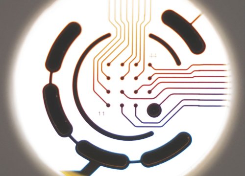
Recordings can be interpreted as local field potentials (LFPs), or EEG-like signals, for detection and amplification of activity. Measuring LFPs allow for continuous streaming of data from multiple electrodes surrounding the larva. Prior to solidification of the low melt agarose, larva were positioned with the dorsal side of their head and body in contact with electrodes. Once the agarose solidifies, recordings of spontaneous activity were taken over a 10-minute period. Larva need to be completely immobilized in contact with the electrodes as activity is not observed if the larva is moving. Axion's Maestro MEA systems provide great sensitivity to detect and process these signals. Here is the snapshot of activity noting relative head or brain LFP rate in relative amplitude or more posterior for relative spinal cord activity. We overall observe diminished brain activity after valproic acid treatment.
Significantly decreased activity was identified in the average number of LFPs, mean LFP rate, and inter-LFP interval coefficient of variation after treatment with 500 uM valproic acid compared to control. The average number, frequency and percentage of electrographic burst events, along with the LFPs per electrographic burst, were significantly decreased after treatment. No difference was observed in electrographic burst duration or an inter-electrographic burst event properties, indicating valproic acid decreased LFP propensity but not overt LFP properties. This coincides with reported valproic acid action on glutamatergic excitatory neurons. We were additionally able to measure relative network activity across the brain. Overall we did not observe broad changes to network activity but there was a significant decrease in the number of LFPs per network electrographic burst per channel, and network ILI coefficient of variation, indicating decreased detection of LFPs across the brain yet with less variability.
Another advantage of this MEA system is simultaneous measurement of brain and spinal cord activity. Data was analyzed and pulled for electrodes in contact with the spinal cord region anterior to the swim bladder. Significant differences in activity were not observed between valproic acid treatment, yet we noted overall activity is substantially higher compared to head measurements. This observation of diminished brain activity but not spinal cord after treatment with valproic acid supports brain activity differences or not a secondary effect from decreased locomotion. This accessible method uses the commercially available multi-electrode array to detect local field potential parameters and allows for recording of relative coordinated network activity. we provided a step-by-step protocol for the experiments presented that a novice could easily follow an accessible electrophysiological studies in the neuroscience field. Future direction with this methodology is activity after stimulation in the MEA plate.
Thank you. And that is the conclusion for today's Coffee Break Webinar. If you have any questions you would like to ask regarding the research presented, or if you are interested in presenting your own research with microelectrode array technology or impedance based assays, please forward them to: coffeebreak@axionbio.com
For questions submitted for Dr. Tomasello she will be in touch with you shortly. Thank you for joining in on today's Coffee Break Webinar and we look forward to seeing you again.