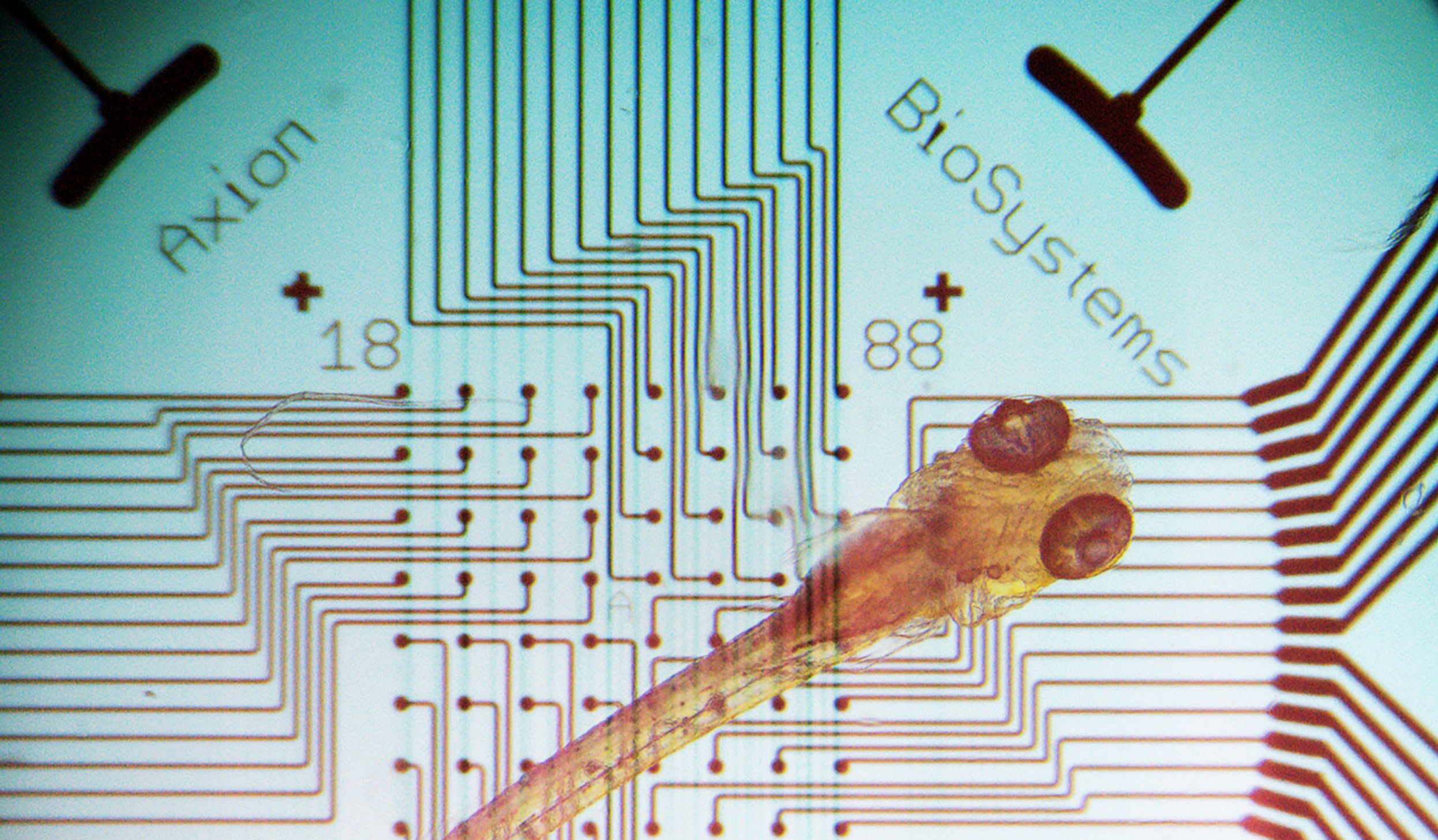
Zebrafish are a great translational model of whole organ systems. Their accessibility, ease of genetic engineering, and simple behavioral screens make them useful models for many diseases. Zebrafish models are often used for toxicology and drug discovery research.
Expand on standard zebrafish assays with noninvasive electrophysiological readouts from the brain and spinal cord. Measure real-time drug interactions in live zebrafish. Axion BioSystems’ multielectrode array (MEA) assays are well-suited for studying zebrafish neurobiology.
Scale up organism electrophysiology
Measuring neural activity in living zebrafish has always been a challenge. Consistently capture zebrafish electrophysiology simultaneously across multiple wells from live zebrafish with MEA. Multiple electrodes measure independently across the central nervous system to evaluate neural networks non-invasively and label-free. Specific regions, like the forebrain, midbrain, or hindbrain, may be targeted based on the zebrafish orientation in the well.
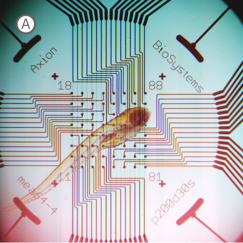
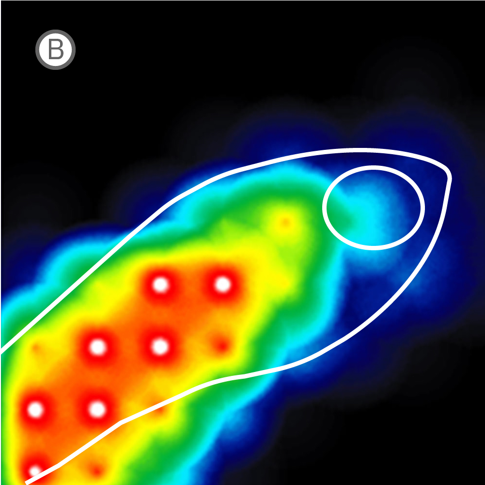
(A) Inverted microscope image of zebrafish larva in a CytoView MEA plate. (B) Neural activity map illustrating that the detected extracellular activity aligns with zebrafish orientation.
The transparent CytoView plates let you identify which electrodes correspond to which region in the spinal cord or brain. With the option to multiplex standard zebrafish imaging assays on CytoView plates after measuring activity, MEA data can easily supplement many workflows. While recording, regions of activity light up bright on the activity map in AxIS Navigator while uncovered electrodes remain dark.
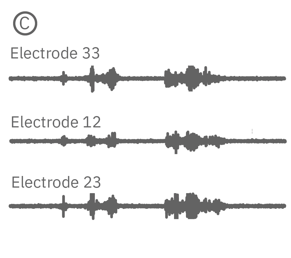
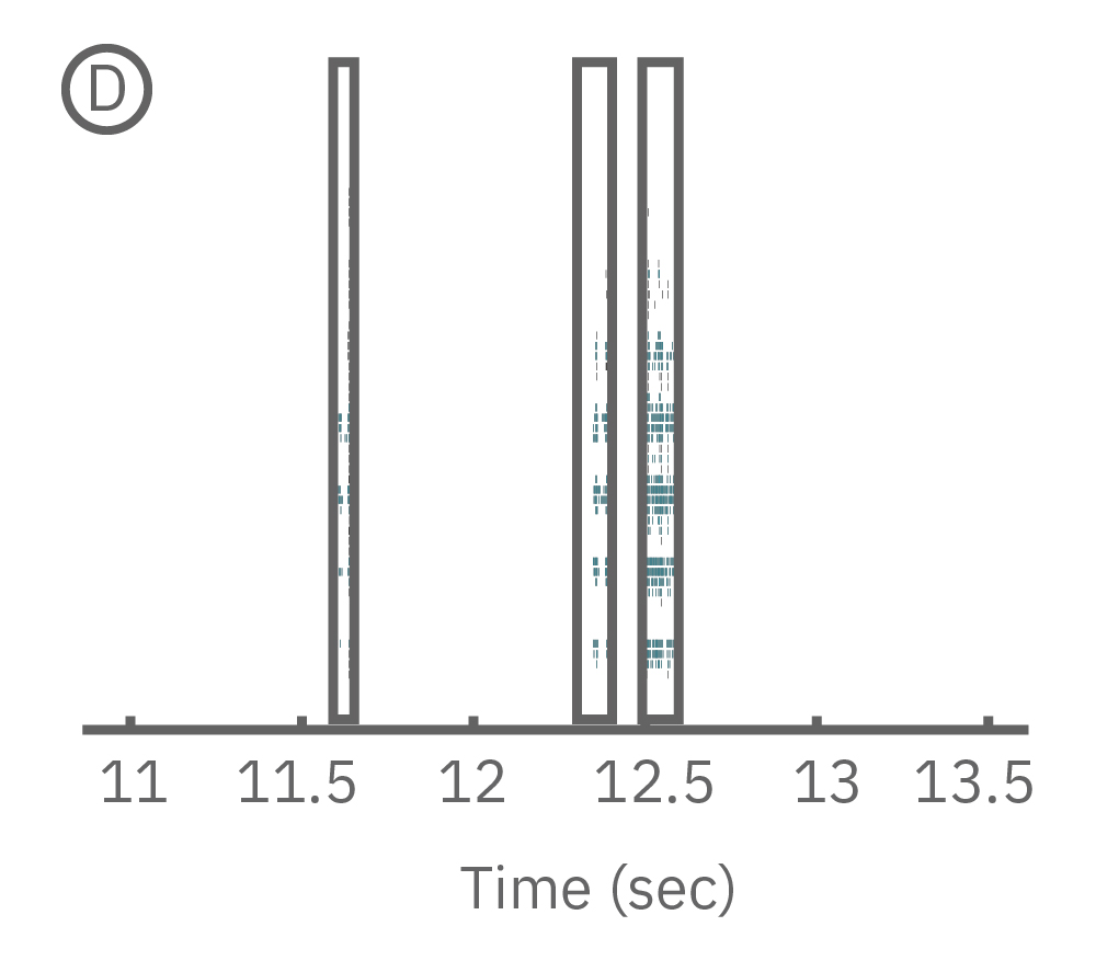
(C) Continuous data recorded from 3 electrodes along a zebrafish spinal cord. (D) Well-wide raster plot demonstrates zebrafish spinal cord activity is organized into synchronous bursts.
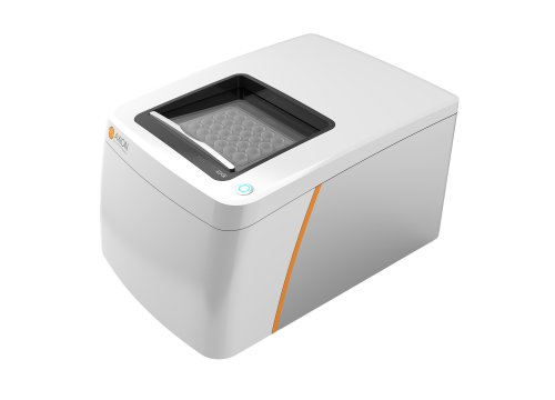
AxIS Navigator lets you analyze spiking events from a single electrode, identify and sort individual neurons, or compare between multiple electrodes; allowing you to analyze localized as well as network or region-wide nervous system activity. Zebrafish electrophysiology has never been easier or more accessible than with the Maestro MEA.
Unlock the potential of your zebrafish models with the Maestro MEA and gain new insights into:
- Neurodevelopmental and degenerative disorders
- Drug or toxicological effects
- Simultaneous brain and spinal cord responses
Read the paper below by Tomasello, 2020 to learn more about the step-by-step process to record electrophysiological data from zebrafish on the Maestro MEA system.
Read Tomasello's Zebrafish Publication
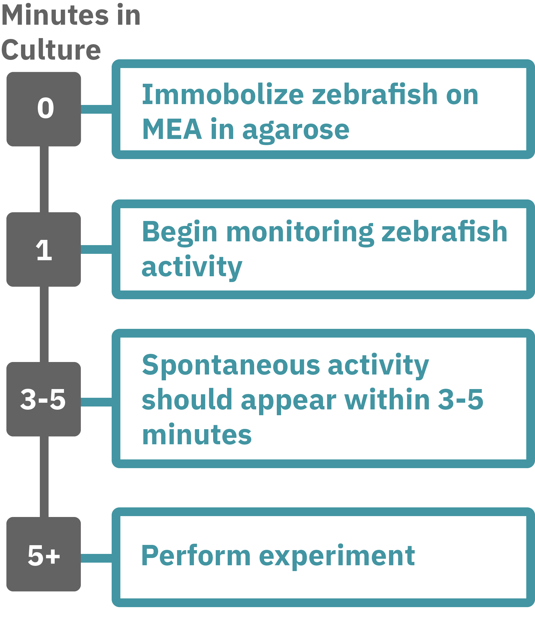
Getting started with Maestro Pro and Edge couldn't be easier. Immobilize the zebrafish in agarose over the electrode containing region of a well in an MEA plate. Once the agarose has solidified, start recording immediately. Spontaneous activity should be detected within 3-5 minutes. Analyze the zebrafish’s neural activity in the MEA plate label-free and in real-time with AxIS Navigator Neural Module software. Add test compounds as required.

The advantage of measuring zebrafish brain and spinal cord electrical activity on the Maestro Pro and Edge systems:
-
Noninvasive measurement from an intact organism – The Maestro MEA platform performs noninvasive electrical measurements from the zebrafish’s neurons, circumventing the use of dyes/reporters that can perturb your cell model and confound results.
-
It's easy – You don't have to be an electrophysiologist to use the Maestro MEA system. Just immobilize your zebrafish in agarose in an MEA plate, load your plate into the Maestro MEA system, and record your neural data. Axion's data analysis tools will do the rest, even generating the publication-ready graphs you need.

Neural MEA
Show Full Details
What is a microelectrode array (MEA)?
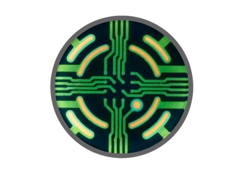
Microelectrode arrays (MEA), also known as multielectrode arrays, contain a grid of tightly spaced electrodes embedded in the culture surface of the well. Electrically active cells, such as neurons, are plated and cultured over the electrodes. When neurons fire action potentials, the electrodes measure the extracellular voltage on a microsecond timescale. As the neurons attach and network with one another, an MEA can simultaneously sample from many locations across the culture to detect propagation and synchronization of neural activity across the cell network.
That’s it, an electrode and your cells. Since the electrodes are extracellular, the recording is noninvasive and does not alter the electrophysiology of the cells - you can measure the activity of your culture for minutes, days, or even months!
Watch the full video and discover if an MEA assay is right for your research.
Watch the full MEA video now

An MEA of 64 electrodes embedded in the substate at the bottom of a well.

Neurons attach to the array and form a network. The microelectrodes detect the action potentials fired as well as their propagation across the network.
Brain waves in a dish
Neurons communicate with other cells via electrochemical signals. Many neural cell types form cellular networks, and MEAs allow us to capture and record the electrical activity that propagates through these networks.
Neurons fire action potentials that are detected by adjacent electrodes as extracellular spikes. As the network matures, neurons often synchronize their electrical activity and may exhibit network bursts, where neurons repeatedly fire groups of spikes over a short period of time.
The MEA detects each cell's activity, as well as the propagation of the activity across the network, with spatial and temporal precision. Patterns as complex as EEG-like waveforms, or "brain waves in a dish", can be observed. Axion's MEA assay captures key features of neural network behavior as functional endpoints - activity, synchrony, and network oscillations.

Action potentials are the defining feature of neuron function. High values indicate frequent action potential firing and low values indicate the neurons may have impaired function.

Synapses are functional connections between neurons. Synchrony reflects the prevalence and strength of synaptic connections, and thus how likely neurons are to generate action potentials simultaneously on millisecond time scales.

Network oscillations, or network bursting, as defined by alternating periods of high and low activity, are a hallmark of functional networks with excitatory and inhibitory neurons. Oscillation is a measure of how the spikes from all of the neurons are organized in time.
Do more with multiwell
Axion BioSystems offers multiwell plates, ranging from 6 to 96 wells, with an MEA embedded in the bottom of each well. Multiwell MEA plates allow you to study complex neural biology in a dish, from a single cell firing to network activity, across many conditions and cell types at once.

Optogenetics
Show Full DetailsOptogenetics: Using light to control cells
Optogenetics is a technique that involves the use of light to control cell function. Cells are first genetically modified to express light-sensitive ion channels, called opsins. Then, light can be used to activate the opsin. The most well-known opsins are light-gated ion channels that can control the excitability of the cell membrane. When activated by the opsin-specific wavelength of light, the channels open allowing ions to flow across the cell membrane to either excite or inhibit the cell. Optogenetics enables precise control over a targeted cell population.
Many opsins, many options
Since the first microbial opsin was introduced into mammalian neurons in 2005, many different opsins have been used to control the excitability of electroactive cells such as neurons and cardiomyocytes. Each opsin is sensitive to a specific wavelength range, or color of light and induces a precise biological event.

Channelrhodopsin (ChR2), for example, is activated by blue light. When ChR2 opens, positive cations (like sodium and calcium), flow into the cell, depolarizing or exciting the cell. In contrast, halorhodopsin and archaerhodopsin both inhibit cell excitability by hyperpolarizing the cell in response to yellow or green light, respectively. With optogenetics, you can turn on and off cells like a light switch.
The timing of these light-activated events is fast, facilitating highly precise control. First generation opsins, such as channelrhodopsin, open and close in milliseconds, ideal for kicking off an action potential. Second generation opsins have fined tuned kinetics for even faster, more precise control or slower, longer-lasting inhibition. For example, step-function opsins stay open until another pulse of light switches them off.


Optogenetics can precisely control which cells are turned on or off by employing different gene promoters for opsin expression. Opsins can be expressed in all neurons or used to control specific subpopulations. Unlike electrical stimulation, which excites all nearby cells, optical stimulation can be finely targeted to the cells expressing the opsins responsive to a narrow band of light wavelengths.


In summary, optogenetics is a powerful toolbox for precise control over targeted cell populations at fast time scales. Superior spatial and temporal control, reversibility, and easy stimulus delivery make exploring complex biology simpler than ever before.
More than just ion channels
As the field has advanced, opsins have been used to control more than just ion flow. Light-activated gene expression with light-inducible transcription factors can control the proteins made by cells. The combination of optogenetics with CRISPR provides even greater control over CRISPR/Cas9 gene editing.
Opsins have also been incorporated into many biochemical and intracellular signaling pathways to control key protein functions. MAPK and PI3K pathways, Rho family GTPase activation, apoptosis, and protein trafficking can now all be precisely controlled by light.

Shining light in vitro
Sophisticated biology like optogenetics demands sophisticated technology to explore it. In vitro technology relied on single wavelength lasers and custom lab-specific tools while many in vivo technologies were quickly developed for optical stimulation. The Lumos Optical Stimulation system is the first-of-its-kind multiwell optical stimulator with the ability to deliver up to four wavelengths of light per well with microsecond precision. From controlling the excitability of your neurons to pacing the beating of your cardiomyocytes, discover how the Lumos and optogenetics can revolutionize your assay.