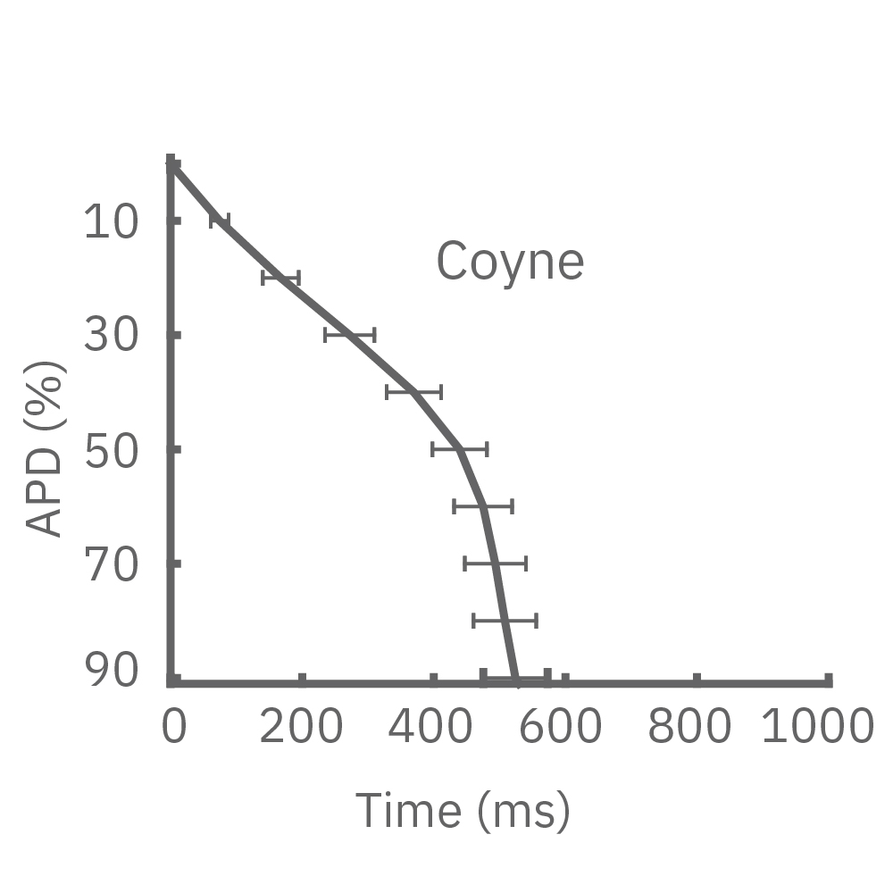
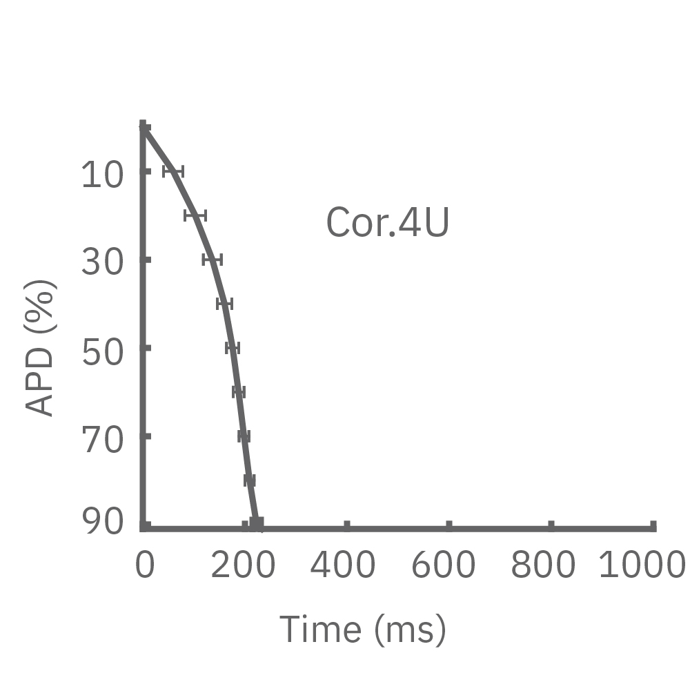
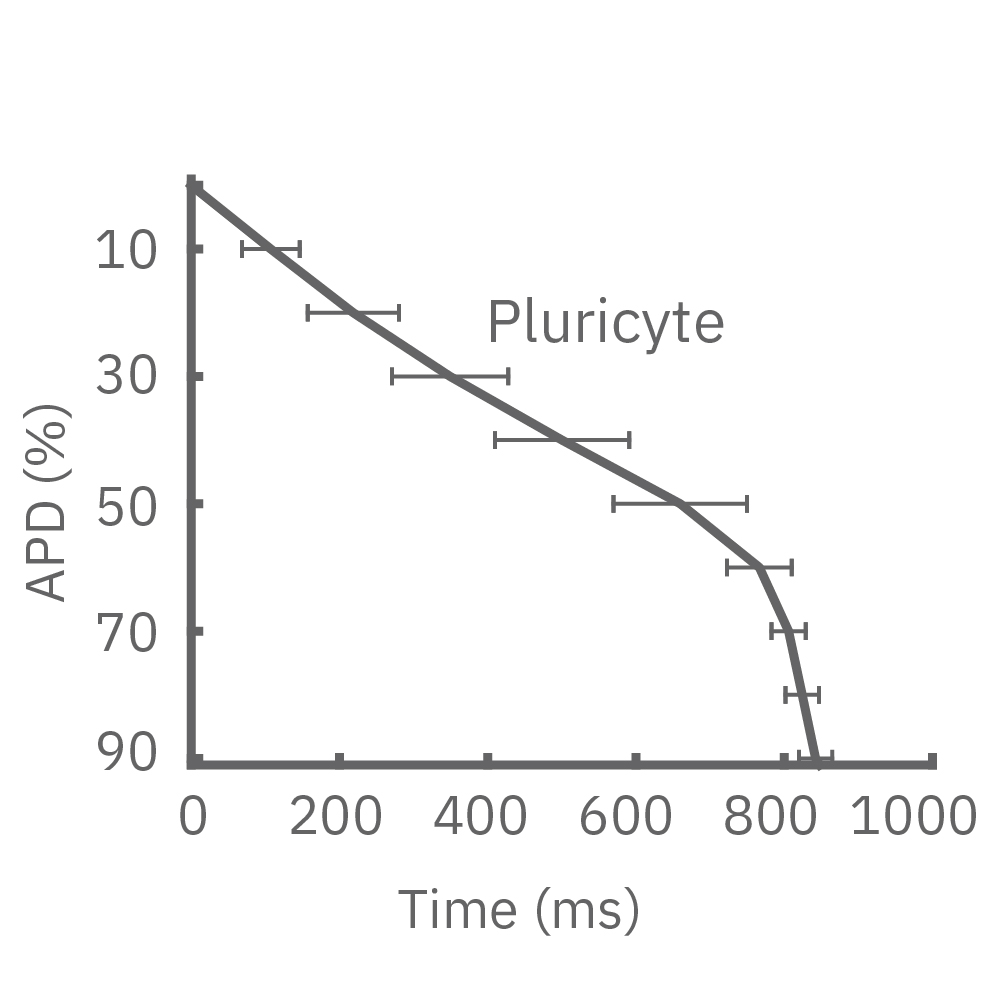
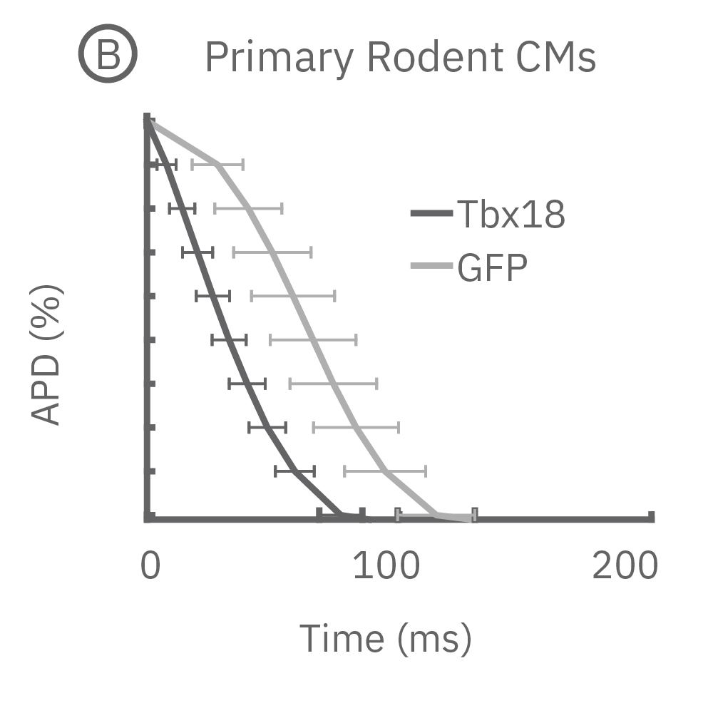

Publication
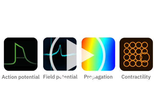
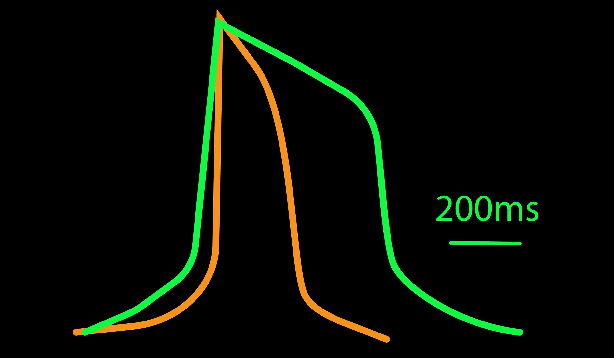
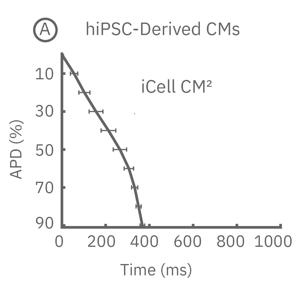
Novel method for action potential measurements from intact cardiac monolayers with multiwell microelectrode array technology
Author:
H. B. Hayes, A. M. Nicolini, C. A. Arrowood, S. A. Chvatal, D. W. Wolfson, H. C. Cho, D. D. Sullivan, J. Chal, B. Fermini, M. Clements, J. D. Ross, and D. C. Millard
Product:
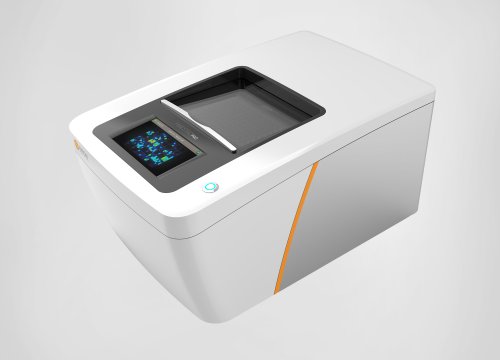
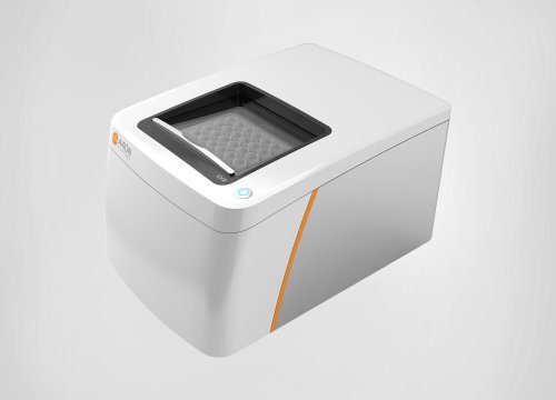
Maestro Pro,
心筋モジュール,
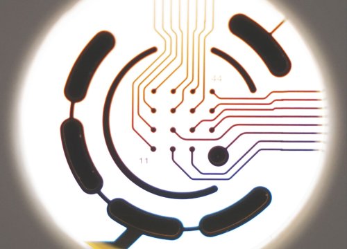
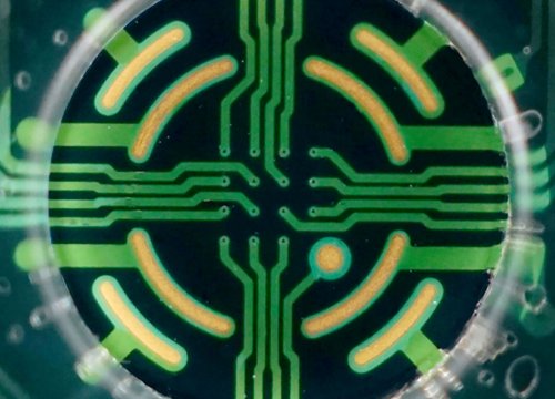
BioCircuit MEA プレート,