
Coffee Break Webinar
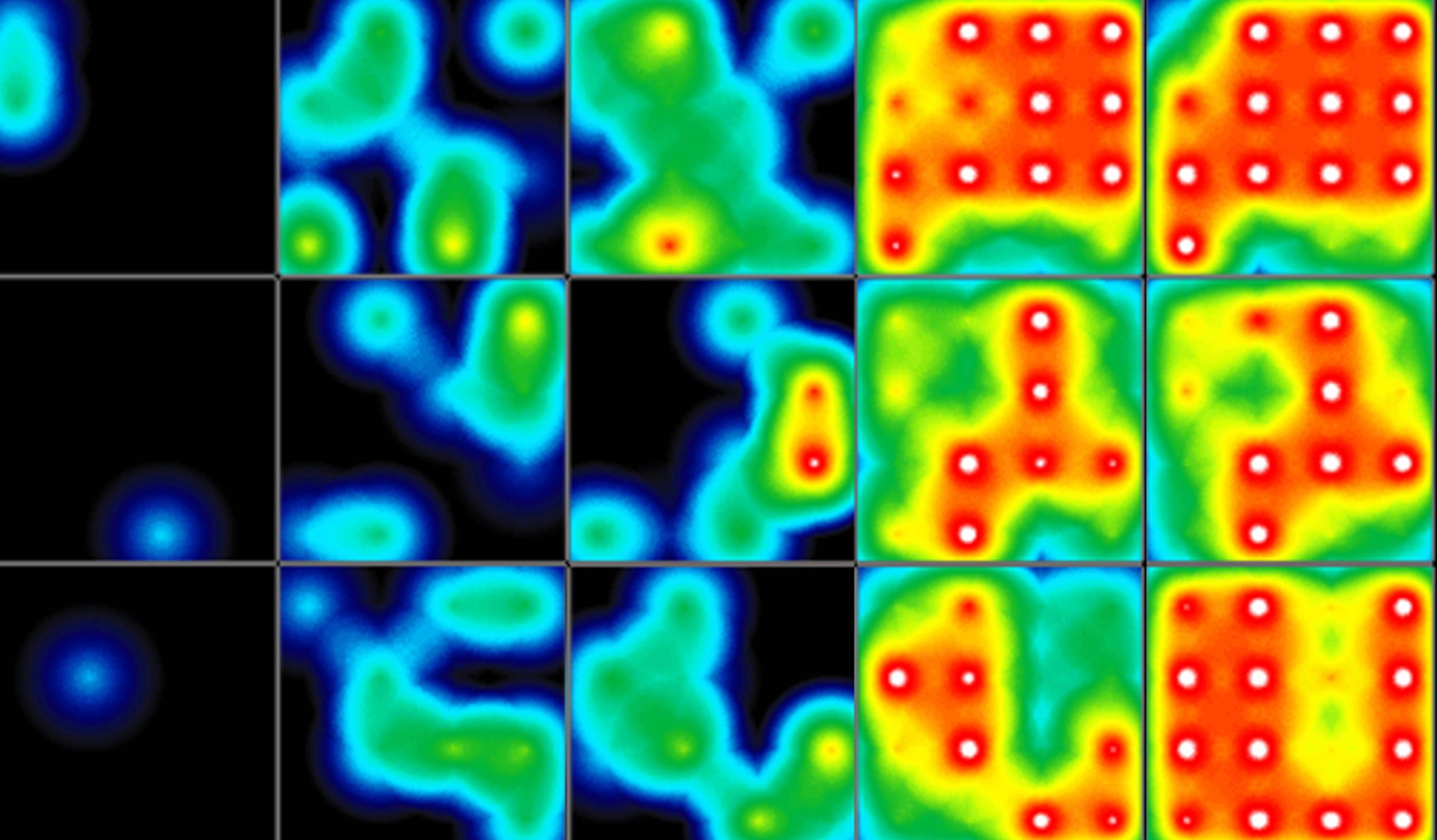
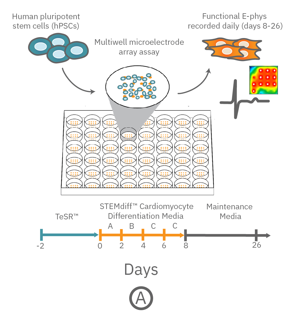
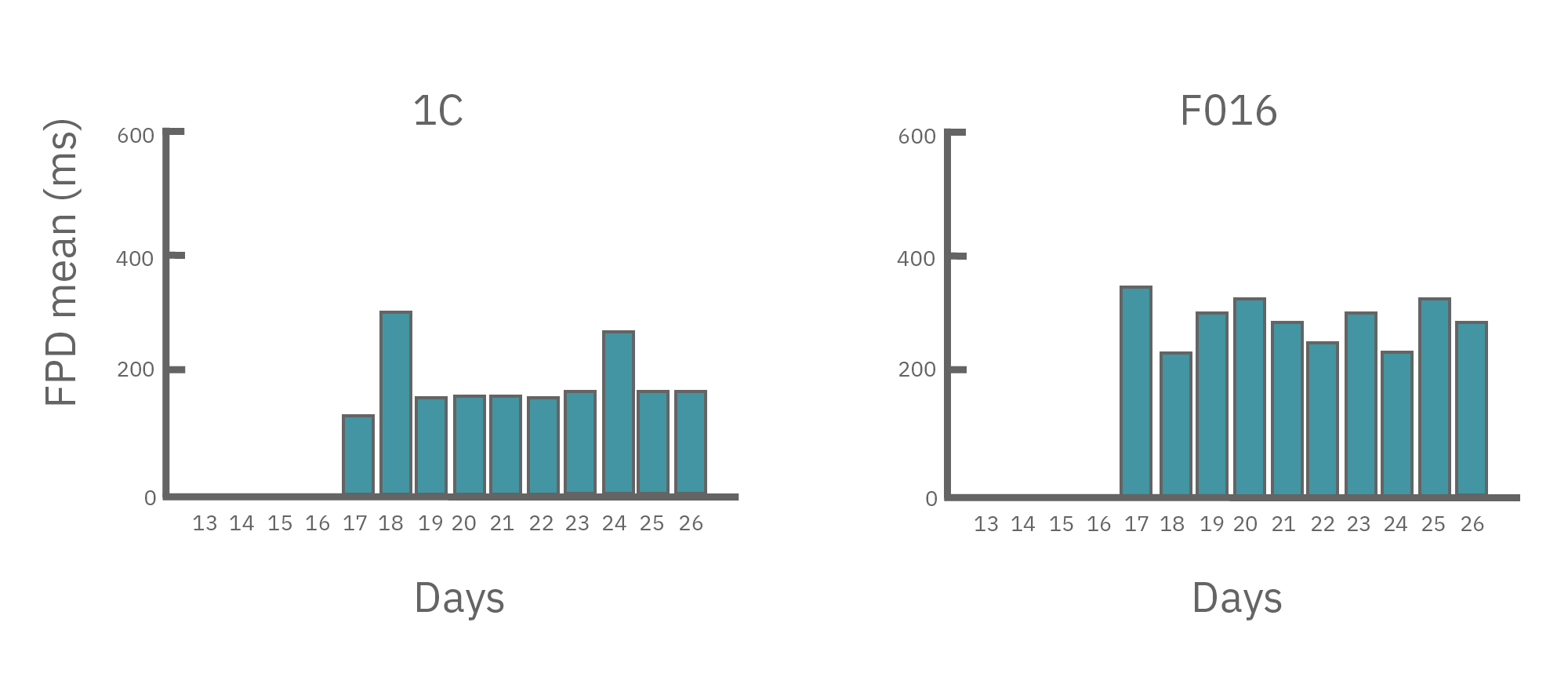
Recreating irregular heart beats with patient cells and gene editing
Author:
Dr. Vincenzo Macri
Product:
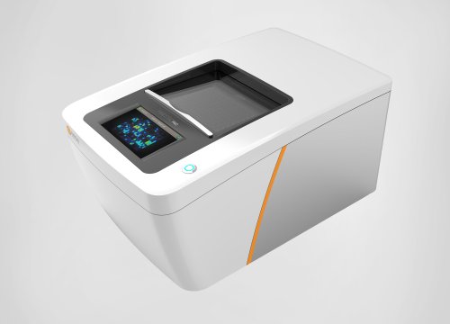
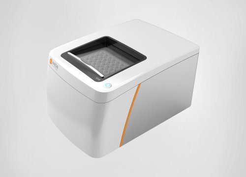
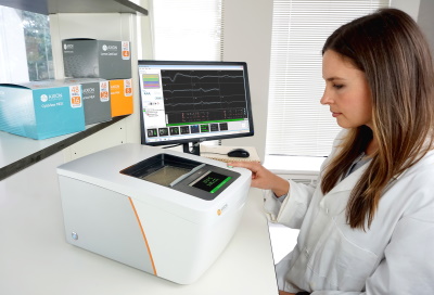
Maestro Pro,
心筋モジュール,
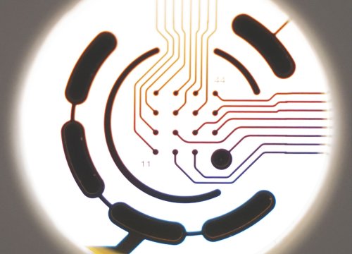
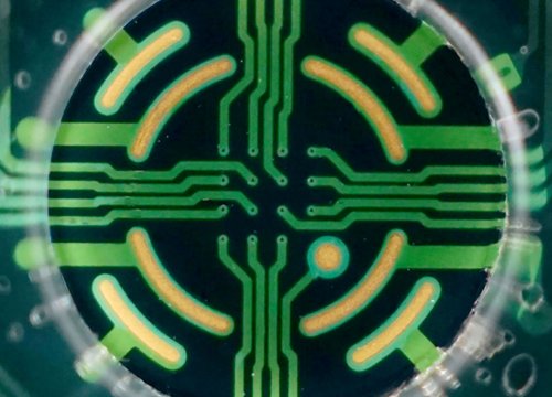
CytoView MEA プレート,
Maestro Edge,