Publication
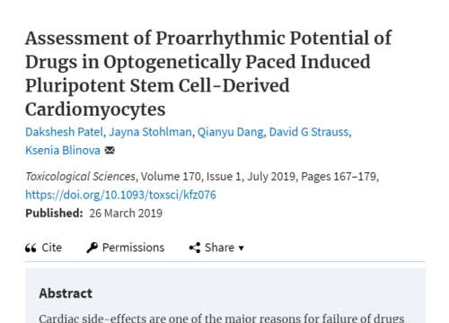
Assessment of proarrhythmic potential of drugs in optogenetically paced induced pluripotent stem cell-derived cardiomyocytes
Author:
Patel D, Stohlman J, Dang Q, Strauss DG, Blinova K.
Product:
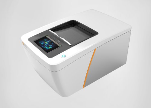
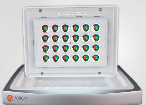
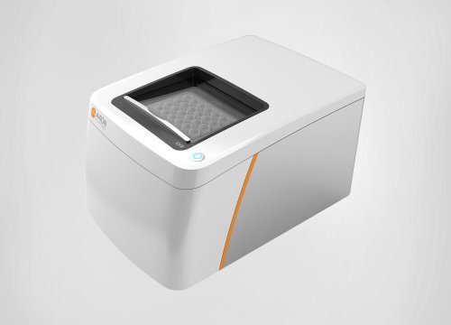
Maestro Pro,
心筋モジュール,
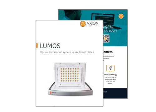
Lumos MEA プレート,
Lumos,
Maestro Edge,