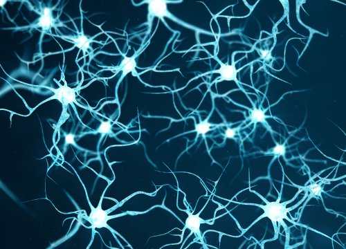
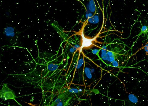
神経共培養とグリア細胞相互作用
異種細胞と共培養された神経共培養。培養内の細胞組成改変により、神経発達、疾患進行などを検証します
Learn More→
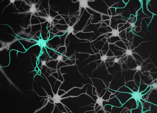
Neural MEA (microelectrode array) technology enables noninvasive, real-time recording of electrical activity across neuronal networks cultured on embedded electrode arrays. By capturing action potentials and network-level firing patterns over time, MEA assays provide functional insight into neural health, connectivity, and drug response.
Traditional neuroscience assays often rely on indirect proxies or low-throughput single-cell techniques. Neural MEA monitoring delivers direct electrophysiological readouts across many wells simultaneously, enabling scalable discovery workflows while preserving neuronal biology.
![]() Label-free, noninvasive recording of spontaneous and evoked neural activity
Label-free, noninvasive recording of spontaneous and evoked neural activity
![]() Network-level functional readouts including firing rate, bursting, and synchrony
Network-level functional readouts including firing rate, bursting, and synchrony
![]() Longitudinal monitoring over days to weeks in stable culture conditions
Longitudinal monitoring over days to weeks in stable culture conditions
![]() Multiwell throughput compatible with drug discovery and toxicity screening
Multiwell throughput compatible with drug discovery and toxicity screening
![]() Quantitative, reproducible electrophysiology data that provide a robust descriptor of neural activity
Quantitative, reproducible electrophysiology data that provide a robust descriptor of neural activity
![]() Compatible with both 2D monolayer cultures and 3D models (e.g., spheroids and organoids), enabling physiologically relevant experimental design
Compatible with both 2D monolayer cultures and 3D models (e.g., spheroids and organoids), enabling physiologically relevant experimental design
The Axion BioSystems Maestro Pro has been an exceptional tool for studying electrical signaling data on neurons. Its high-throughput capabilities and user-friendly interface make it ideal for capturing detailed electrophysiological activity.
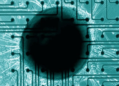
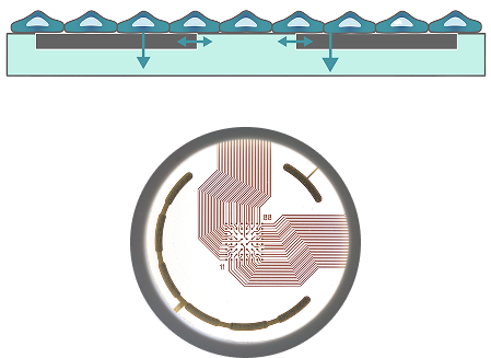
Neurons generate extracellular voltage changes as they fire action potentials. Microelectrodes embedded in the culture surface detect these signals across the network, enabling quantitative assessment of functional connectivity and dynamic response to stimuli or compounds.
Neural MEA workflows integrate seamlessly into standard neuronal culture protocols, enabling straightforward setup with rich functional data output. MEA enables everything from acute assays to longitudinal studies.
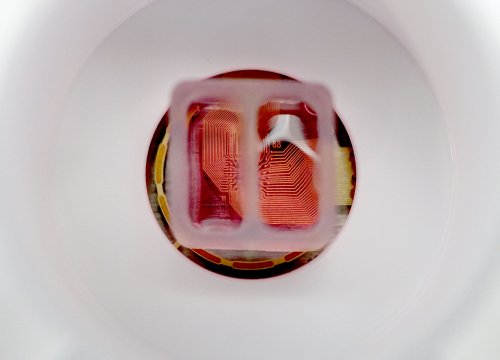
Neurons are cultured and seeded onto electrode-integrated multiwell plates using standard coating and plating protocols. No labels or dyes are required.

Plates are placed into the Maestro MEA recording system, which maintains stable environmental conditions and begins extracellular signal acquisition.
Electrical activity is recorded continuously or at scheduled intervals as neuronal networks mature, burst, synchronize, or respond to treatments.

Recorded signals are processed into quantitative metrics such as firing rate, burst structure, synchrony, and connectivity to support neuroscience decision-making.
Explore how Maestro Neural MEA compares to patch clamp, calcium imaging, and HD-MEA platforms for scalable neuroscience research.
Patch clamp provides high-resolution single-cell recordings but is labor-intensive and low throughput. Neural MEA enables parallel, network-level electrophysiology across many wells, supporting scalable functional screening.
Calcium imaging is an indirect proxy for electrical activity and requires fluorescent indicators that can introduce phototoxicity. MEA provides direct, label-free electrophysiological measurements with superior temporal resolution.
HD-MEA offers very high electrode density but often involves complex workflows and limited scalability. Maestro Neural MEA balances robust network resolution with multiwell throughput, standardized analysis, and ease of use for routine screening and disease modeling.
MEA assays quantify firing rate, bursting, synchrony, oscillatory patterns, and functional connectivity, providing deep insight into network development and pharmacological modulation. The full list of neural metrics provided by the Maestro MEA Neural Module can be found here.
Yes. MEA supports longitudinal recording over days to weeks, making them ideal for chronic exposure studies, neurodevelopmental models, and neurodegeneration research.
Maestro MEA does not require extensive electrophysiology training. Installation and user training are typically completed in a single day, and the integrated workflow enables routine recordings and analysis with standard cell culture experience.