By: Daniel Millard, Director of Applications at Axion BioSystems
Immunotherapies show promise, but they’re a long way from perfection. Immunotherapies supercharge the immune system by helping it target and kill tumor cells. This class of treatments is approved to treat at least 20 different cancers today, but only 15–20% of those who receive immunotherapy show a durable response and side effects are not uncommon. Due to the complexity of the immunotherapy manufacturing process, especially for adoptive cell therapies such as CAR-T cell therapy, immunotherapy potency is hard to predict. As scientists seek to develop safer, more effective immunotherapies and expand their reach to treat additional cancer types, ensuring the consistency of cell-based products is critical—and so is choosing the right cell potency assay.
A potency assay should quantify the key biological activity of a cellular product. For immunotherapies, the key biological process is the ability of immune cells to target and kill cancer cells. Researchers use cell viability and cytotoxicity assays to measure how swiftly and completely their treatments kill tumor cells in vitro, creating a benchmark to gauge performance between batches of manufactured products. This quantitative approach to assessing potency helps ensure consistency with the goal of improving therapeutic outcomes.
Scientists can go about testing cytotoxicity in several different ways. Here are five commonly used cell viability assays and their benefits and limitations in immunotherapy potency testing.
1. Chromium release assays
The chromium release assay is a widely accepted cell viability assay. Since 1968, it has been the most used method for assessing the cytotoxicity of adoptive cell therapies such as CAR-T cells. Researchers load target cells, such as tumor cells, with a radioactive isotope of chromium and then expose the cells to active CAR-T or CAR-NK cells. After an incubation period, researchers probe the cell culture medium for a radioactive signal. The strength of this signal is directly proportional to the number of target cells destroyed.
Although this assay has become commonplace, some labs are shifting away from it in favor of more powerful and sensitive assays. Chromium release assays are limited by their nature as an endpoint method. Researchers can only assess cell killing at one point in time, meaning they cannot measure the kinetics of cytotoxicity to determine the peak killing rate, signs of chronic activation, or the point of T-cell exhaustion. Furthermore, this test uses harmful radiation. New assays use different types of signals that are less toxic to users.
2. MTT assays
Researchers can use the tetrazolium salt MTT as an indicator of mitochondrial metabolic rate, a proxy for cell viability. If functional, mitochondrial enzymes cause MTT to form into purple crystals that researchers can measure using spectrophotometers. The more intense the purple color, the higher the number of viable cells in the medium.
Many researchers use this method to assess the cytotoxicity of immunotherapies. It is inexpensive and easy to perform, and unlike chromium release assays, it does not use radiation. But like chromium release assays, MTT assays are endpoint reactions, limiting the amount of information a researcher can collect about the cell-killing behavior of immunotherapies.
3. LDH assays
Lactate dehydrogenase (LDH) is present in all cells, so the chemical leaks out when an immunotherapy treatment damages a cell membrane. Accordingly, the concentration of LDH in a cell culture medium indicates the magnitude of a treatment’s cytotoxicity.
This assay is an effective method for determining the potency of immunotherapies but requires a complex protocol. Researchers measure LDH levels by quantifying an index of its activity. LDH converts lactate into pyruvate, which then interacts with a tetrazolium salt to form a water-soluble, red molecule that researchers can detect using colorimetry. The concentration of this red color correlates with the level of cell death.
Like chromium release and MTT assays, LDH assays only measure cell death at a single time point. This limitation makes it difficult to assess the kinetics of a therapy’s activity and produces fewer data for potency calculations.
4. Live-cell imaging
Of all the methods described so far, live-cell imaging is the first to measure cell-killing in real-time. Researchers can monitor their cells continuously over days to months. This capability is ideal in situations where it is difficult to predict the timing of certain events, such as the kinetics of CAR-T cell activity. Yet live-cell imaging presents its own limitations.
Live-cell imaging uses optical techniques to monitor cells in culture. When testing adoptive cell therapies, researchers plate tumor cells and place engineered immune cells on top of them. But the immune cells can block the view of the tumor cells, making it challenging to measure cell killing reliably. Scientists can use dyes to differentiate the two cell populations, but this additional step complicates the protocol and risks affecting cells’ natural behavior. Also, since the data are visual, data analysis is subjective and requires a trained eye to interpret the results.
5. Bioelectronic assays for impedance monitoring
The latest generation of cell viability assays includes bioelectronic assays, which solve many of the challenges inherent to prior cell viability assays. Bioelectronic assays enable real-time, quantitative measurement of the immune cell-mediated killing of tumor cells without using dyes or labels that can interfere with cell behavior.
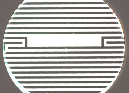
Bioelectronic assays start with a multiwell plate that contains bioelectronic sensors at the bottom of each well. Researchers plate tumor cells, which attach to the electrodes and impede the electrical signal. Next, researchers add the immune cells and watch as they kill the tumor cells. As tumor cells die and detach, the recorded impedance goes down. This non-invasive technique enables researchers to quantitatively measure cell death in real time over days, weeks or months. Researchers can monitor the cell death kinetics and magnitude across a range of cell concentrations simultaneously to help researchers identify the therapy's potency. Dr. Lohitash Karumbaiah, an associate professor at the University of Georgia, used this technique to assess the potency of a novel CAR-T cell therapy against glioblastoma cells.
Measuring cell viability to assess immune cell potency
Cell viability assays, especially those that enable real-time monitoring of immune cell-tumor interactions, give researchers a reliable method for analyzing the potency of novel immunotherapy candidates. Bioelectronic assays offer the best of all worlds by enabling quantitative, non-invasive, continuous monitoring of immune cell dynamics. The data collected with these impedance assays can help developers screen out ineffective and toxic biotherapeutic candidates during the early development process and reduce the likelihood that subpar treatments will leave the laboratory during manufacturing. In the long term, bioelectronic assays will help bring safer, more effective therapies to patients in need.