CytoView MEA Plate
CytoView MEA 플레이트는 투명한 바닥면을 통해 세포 시각화 및 다중 실험 수행 그리고 미세전극에 의한 세포의 전기적 네트워크 정보와 같은 서로 비교 불가능한 데이터를 동시에 확보할 수 있도록 고안된 최상의 Maestro multiwell MEA 플레이트 입니다.
6,24, 48 그리고 96-well 플레이트를 제공하고 있으며 낮은 잡음 신호와 각 well 당 업계 최고로 많은 수의 미세 전극을 포함하고 있어 며칠 혹은 몇 개월의 데이터 측정이 가능합니다.
Key Features
매 실험에서 더 많은 데이터를 확보하세요.
-
높은 수준의 데이터를 확보 - 심근 세포 혹은 신경 세포 실험에서 보다 상세한 데이터를 획득하기 위해 업계 최고로 각 well 당 많은 미세 전극 수를 제공합니다. PEDOT 전극 기술은 최고 수준의 전기 신호 수집을 보장합니다.
-
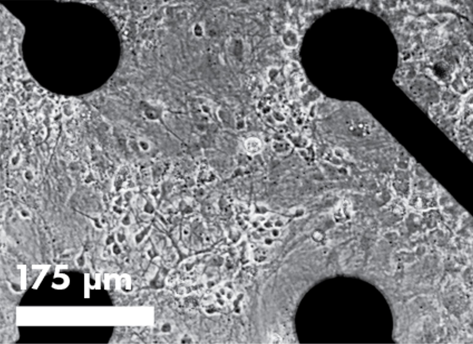
투명한 플레이트 바닥으로 세포 직접 관찰 - 세포 배양 상태를 명시야 현미경을 통해 매일 모니터링 할 수 있습니다.
-
MEA플레이트에서 다양한 실험 결과 확보 - 형광 혹은 발광 플레이트 리더들을 사용하여 다양한 실험을 수행할 수 있습니다. 최적의 실험 결과 획득을 위해 검은색 혹은 흰색의 플레이트를 선택할 수 있습니다.
CytoView MEAThe CytoView MEA plates combine robust data collection with a transparent well bottom for cell visualization and assay multiplexing
|
||||||||||
| Plate | Cat No. | Wells |
well |
layout* |
|
|
Edge |
Pro |
Z/ZHT |
Original |
|---|---|---|---|---|---|---|---|---|---|---|
| CytoView MEA 6 | (a) M384-tMEA-6B (b) M384-tMEA-6W |
|
PEDOT |
 |
|
(b) White |
|
|
|
|
| CytoView MEA 24 | M384-tMEA-24W |
|
PEDOT |
 |
|
|
|
|
|
|
| CytoView MEA 48 | (a) M768-tMEA-48B (b) M768-tMEA-48W |
|
PEDOT |
 |
|
(b) White |
|
|
||
| CytoView MEA 96 | (a) M768-tMEA-96B (b) M768-tMEA-96W |
|
PEDOT |
 |
|
(b) White |
|
|
||
*Schematic of well illustrating recording electrodes (blue), grounds (orange), and where present, a large dedicated stimulation (blue).
Overview
세포 이미징 및 여러 실험 동시 수행
혁신적인 투명한 플레이트 바닥은 세포 시각화 및 여러 실험을 동시 수행할 수 있도록 지원합니다. 명시야 이미징은 세포가 정확한 위치에 자리잡았는지 확인할 수 있도록 도와주고 세포가 건강하게 서로 연결되었을 때 보여지는 MEA 결과에 신뢰를 더해줍니다. 또한 여러 형광 혹은 발광 실험과 같은 End point 실험 결과로 MEA 측정 결과를 보완할 수 있습니다.
높은 수준의 MEA 데이터 획득
심장세포와 신경세포 모두 Cytoview MEA 플레이트에서 매우 잘 자라며 높은 신호대잡음비(Signal-to-Noise) 를 보여줍니다.
심장세포와 신경세포 모두 Cytoview MEA 플레이트에서 매우 잘 자라며 높은 신호대잡음비(Signal-to-Noise) 를 보여줍니다
투명한CytoView MEA 플레이트의 동일 well 에서 리포터 기반 실험(형광 혹은 발광)을 통해 획득한 결과는 MEA 데이터의 신뢰도를 높이는 데에 기여합니다. MEA 데이터와 리포터 기반 실험 결과의 조합은 복합적으로 작용하는 세포 기전을 이해하는 데에 도움이 되는 정보를 제공할 수 있습니다.
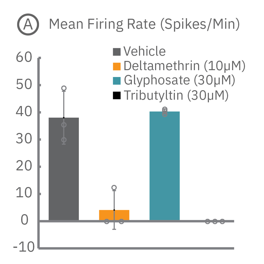
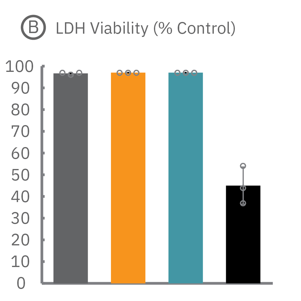
In this neurotoxicity assay, compound were assessed using MEA electrophysiology data from Axion’s Maestro MEA system multiplexed with a fluorescence lactate dehydrogenase (LDH) cell viability assay. Multiplexing enabled greater specificity for compound classification compared to MEA data alone. (A) Both deltamethrin and tributyltin reduced mean firing rate, but (B) only tributyltin reduced cell viability. (Data provided by external customer)