신경 오가노이드와 스페로이드는 인간 뇌의 생리학적, 구조적 그리고 발달 과정이 축소된 형태로 생체 외 환경에서 3차 구조로 배양된 세포 유기체입니다. 전체 뇌 구조와 전뇌, 대뇌, 척수와 같은 일부 특정 형태의 오가노이드의 활용이 신경 발달, 질병의 진행 그리고 약물의 안전성 확인 실험 등에 광범위하게 적용되며 동물 모델과 임상 연구 사이의 간극을 극복하는 데 도움이 되고 있습니다.
신경오가노이드에 대한 완벽한 실험 전개
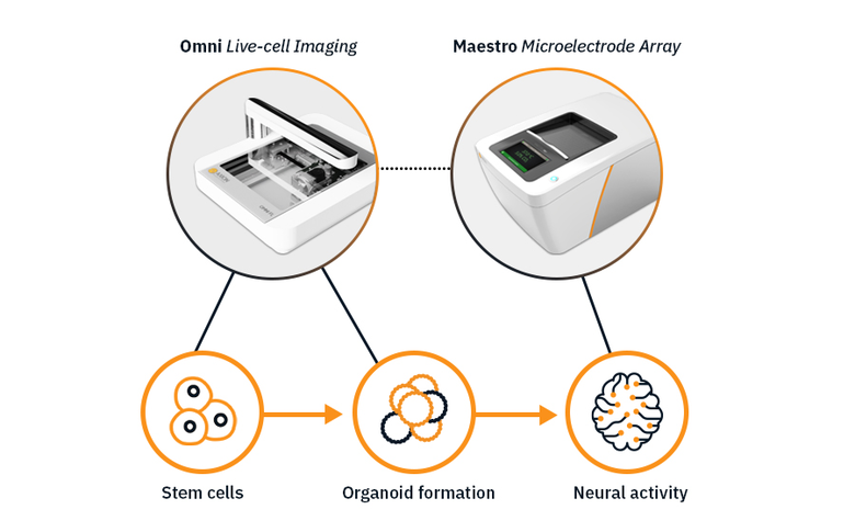
플랫폼의 결합으로 오가노이드 연구 시작부터 마무리까지 수행
좋은 데이터는 안정된 오가노이드로부터 시작됩니다. 줄기세포 배양을 통한 오가노이드 형성 과정을 Omni 를 통해 확인하고 Maestro MEA 를 활용하여 신경 네트워크 발달과 성숙 과정을 관찰해 보세요.
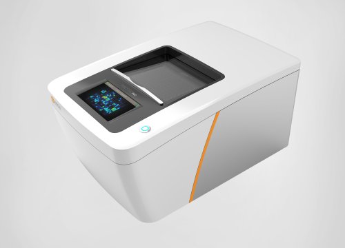
Omni 생세포 이미지 플렛폼
- 줄기세포 배양 과정 모니터링
- 오가노이드 형성 및 생장 추적 관찰
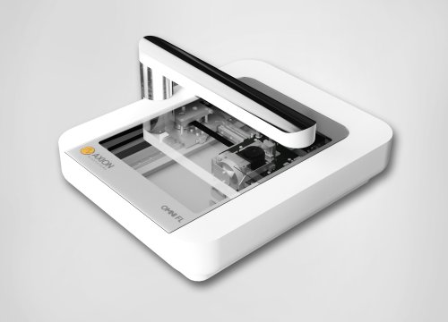
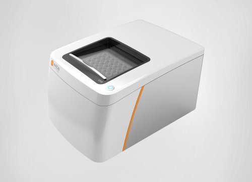
Maestro MEA 플랫폼
- 신경의 활성 기록
- 신경 네트워크와 국소 장 전위(LFP) 발달 정도 측정
신경 오가노이드의 기능적 분석
-
신경 스파이크와 국소 장 전위(LFP)의 동시 측정>
-
Study the development and maturation of brain networks >
-
Develop models of neuroimmune organoid network development>
-
Neanderthal mini-brains>
-
Recording functional activity from cerebral organoids>
-
Assay Steps: Plating neural organoids for measuring activity >
목적: 신경 오가노이드에서 신경 스파이크와 국소 장 전위(LFP)를 동시에 측정한다. 오가노이드는 노의 복잡한 구조와 네트워킹의 축소판으로 발달 과정에서의 LFP 측정이 가능한지 확인.

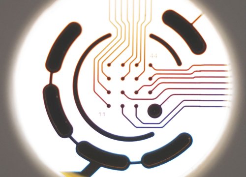
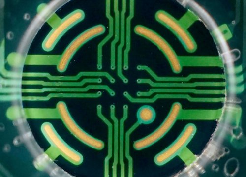
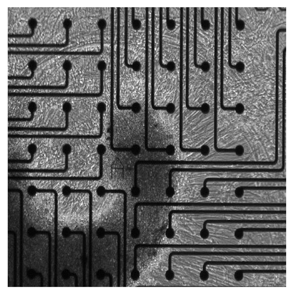
신경 오가노이드를 Maestro MEA 플랫폼으로 측정한 데이터. MEA Viability 모듈로 오가노이드가 MEA Plate 에 잘 부착되었는 지 확인했습니다.
결과: 신경 스파이크가 측면에서만 관측되었고 국소 장 전위(LFP) 는 오가노이드 내측에서 관측되었으며 오가노이드 표면에서는 스파이크가 측정되지 않았습니다.
Purpose: To model human brain development with cortical organoids. Altered brain development in utero can have lifelong consequences but a lack of appropriate in vitro models limits our ability to investigate these processes.
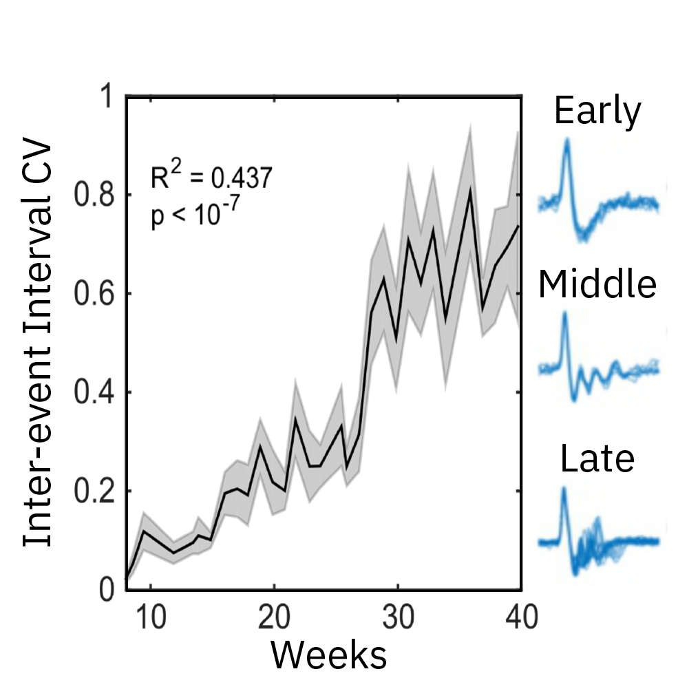
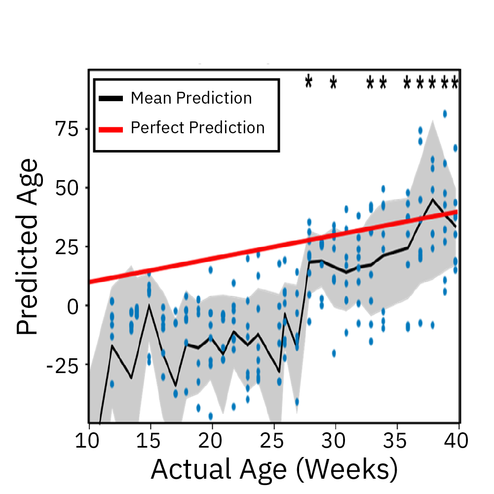
Extracellular recordings of spontaneous electrical activity of neural organoids were measured using the Maestro MEA platform over the course of 10 months and compared to neonatal EEGs.
Result: Cortical organoid activity demonstrated a continuously evolving neural network growing in complexity from early, middle and late time points. Organoid maturation and development were found to mimic the development of the preterm neonatal brain. Watch this webinar to learn more.
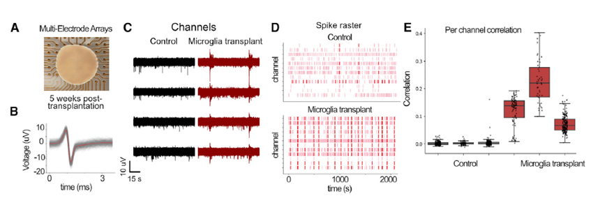
Three-dimensional neural organoid models provide a promising platform for investigating the impact of glial cells on neural activity and exploring their complex role in the pathogenesis and progression of neurological and neuropsychiatric disease. In Popova et al, Cell Stem Cell 2021, the authors compared human microglia across culture models and used the Maestro Pro MEA platform to demonstrate how transplanted microglia accelerated the emergence of synchronous, oscillatory network activity in cortical organoids in vitro.
Cortical organoids on multiwell Maestro MEA system
The genomes of Neanderthals and modern humans are very similar. By investigating the differences in genetic make-up, we can gain insights into what separated modern humans from our extinct relatives. In this webinar, Cleber Trujillo, PhD (StemoniX) discusses how he introduced an archaic variant gene, NOVA1, into human pluripotent stem cell-derived brain organoids and evaluated the impact on neural activity as recorded on the Maestro MEA system.
Electrical activity is measured from organoids cultured on electrodes. Cerebral organoids generated from human induced pluripotent stem cells (hiPSCs) exhibit spontaneous neural activity, with increasing complexity as networks mature. Since the microelectrode array is two dimensional, signals recorded reflect the activity of the neurons on the bottom of the organoid. For the best results, ensure contact between the electrodes and the bottom of the organoid.
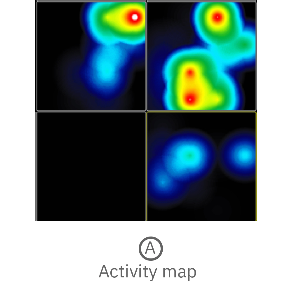
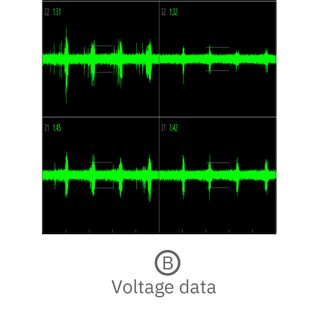
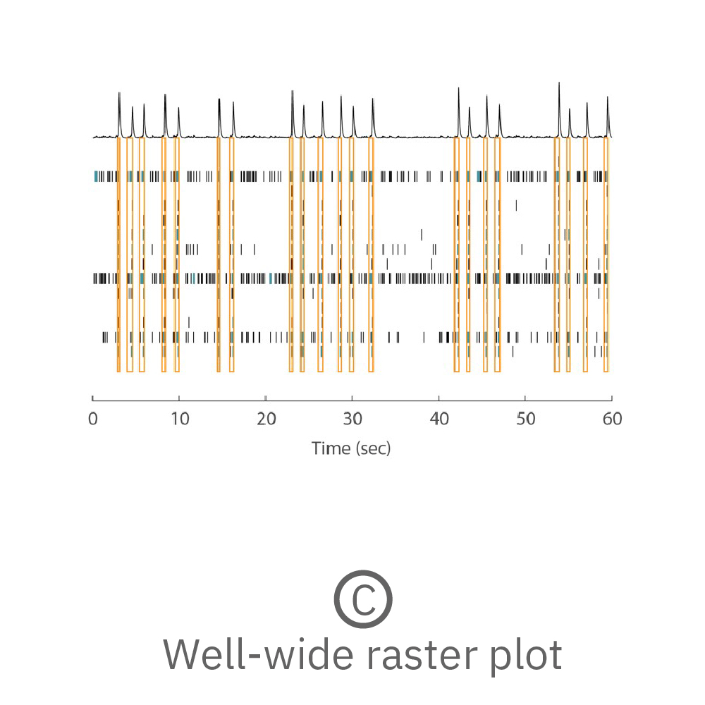
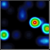
(A) Activity map displaying instantaneous firing rate of four wells with multiple cerebral organoids in each well. (B) Continuous voltage data recorded from four electrodes in one well. Activity is recorded from different sites on the same organoid. (C) Well-wide raster plot showing activity across all 16 electrodes and synchronous network bursting. Teal tick marks indicate electrode bursts, and orange boxes indicate network bursts. Data provided by Maestro customer.
Whole neural organoid plating for MEA recording
This protocol describes a method for plating whole neural organoids on an MEA for measurement of spike and LFP activity.
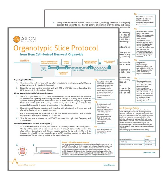
Plating of sliced neural organoids for MEA recording
This protocol describes a method for creating organoid slices for plating on an MEA for measurement of spike and LFP activity.
Neural Organoid Publications and Resources
Neural organoids are rapidly changing the field of neuroscience. See how researchers are using Axion’s live-cell analysis tools to accelerate their organoid research with publications, webinars, application notes, and more.