By: David Ferrick, Chief Scientific Officer of Axion BioSystems
The Pathologist, 2022
Organoids are an increasing presence in the lab – but their full potential will emerge as the technology to understand them evolves.
Behind every medical advancement lies years of exhaustive laboratory research. Scientists rely on a variety of in vitro models to understand human development, investigate disease, and identify therapeutic targets. As novel technologies improve and expand the validation of relevant, “higher-order” in vivo biological models, researchers are performing more in-depth experiments than ever before. For example, mini-organs, or “organoids,” have proven over the past decade to be a valuable resource due to their high biological relevancy – and multielectrode arrays (MEAs) have allowed scientists to decode the range of information they provide.
Organoids are three-dimensional cultures composed of multiple cell types derived from stem cells that self-organize to create miniaturized versions of organs or tissues. To date, the successful generation of numerous mini-organs is seemingly boundless; scientists have created organoids representing the brain, intestine, retina, liver, pancreas, lung, bone marrow, cartilage, breast, prostate, and vascular networks (1). Among these, the most complex model by far is the cerebral organoid, or “mini-brain.”
Cerebral organoids are composed of an intricate network of electroactive neural cells that recapitulate brain activity. Scientists have created mini-brains that mimic a myriad of neurological disorders from autism spectrum disorder (2) to epilepsy (3). But, as scientists identify new uses for cerebral organoids, they must consider the many unique factors involved in recreating neurological conditions in a dish. One of the most important is determining which analytical tools will best capture the data these models can provide.
Neural circuitry made miniature
Since the initial creation of brain organoids, scientists have developed – and are continually refining – protocols to replicate distinct regions, including the cerebellum, thalamus, midbrain, forebrain, and others (4). This specificity has remarkable implications for studying the development and progression of many brain diseases. More recently, scientists have used assembloids – fusions of brain region-specific organoids – to study how circuits form and mature (5). Although these three-dimensional models hold great scientific potential, successful experiments demand robust, data-sufficient technology for assessing neural function.
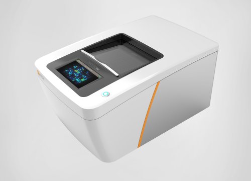
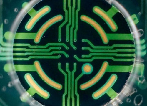
The conventional technique for analyzing the integrated function of neurons is patch-clamp electrophysiology, which is not only technically challenging and highly operator-dependent, but also has very low throughput, because researchers can only examine a handful of cells at a time. This limitation renders patch-clamp electrophysiology unsuitable for measuring the vast network activity organoids produce and is leading to mainstream adoption of MEA-based bioelectronic assays for monitoring organoid function. This type of assay uses a multi-well tissue culture plate holding tightly spaced electrodes at the bottom of each well. These MEAs capture electrical signals emitted by the plated sample, enabling large-scale and real-time measurement of electrical activity in cerebral organoids. Because it does not use dyes, probes, or reagents, the technique does not affect the underlying biology of the sample – and MEA systems are relatively simple to operate without extensive training.
MEAs are long-established for use in assessing two-dimensional samples; now, researchers are finding ways to use them to analyze three-dimensional models. Some dissociate their organoids and plate them for functional assessment; others perform bioelectronic analyses using intact developing organoids to obtain electrophysiological measurements (6). Some data suggest that 2D and 3D cultures display distinct firing patterns (7), highlighting the importance of developing models that closely mimic the complexity of the brain. With bioelectronic assays increasingly used to monitor cerebral organoids, the technique is making 3D applications more accessible than ever (7, 8).
More to see from organoids
Although bioelectronic assays do not require sample visualization, many scientists choose to pair functional analysis using a bioelectronic assay with structural analysis using microscopy. Existing microscopy techniques are optimized for 2D systems, which limits their utility in assessing organoids. In most cases, to image a brain organoid, researchers must slice and fix samples (9). Although this protocol makes it easy for scientists to study cellular composition and morphology, insights from a static tissue slice reveal nothing about how the organoid matures or behaves – and techniques that require scientists to label cells for monitoring can interfere with cell processes and even render the organoid unusable after imaging.
Fortunately, there are some workarounds; for instance, both laser scanning confocal microscopy and two-photon microscopy allow 3D imaging. However, their use is limited by their low image acquisition rate and their potential for phototoxicity and photobleaching. As a result, scientists will often work with a combination of visual strategies to overcome each technique’s individual limitations and improve overall efficiency (10).
Scientists today need solutions for long-term live-cell imaging to better understand organoid development and maximize the amount of information that can be gleaned from these models. The gap is already starting to close as the field begins to pair existing next-generation live-cell imaging technologies with some of the first organoid-dedicated, AI-driven analysis modules. Brightfield live-cell imagers allow users to phenotype organoids and noninvasively track their long-term development. These imagers can measure the diameter, area, number, aspect ratio, and eccentricity of cerebral organoids. It is now also possible to perform live-cell fluorescence microscopy for 3D organoids. The ability to monitor these mini-organs as they undergo maturation will support efforts to fine-tune organoid protocols to minimize variability, improve specification, and advance our understanding of many diseases.
Organoids’ ability to unlock “higher-order” biology depends heavily on the technology available to study them. Fortunately, key techniques like live-cell imaging and bioelectronic analysis can help organoids accelerate personalized medicine by providing more relevant in vivo mimicry and expanding our knowledge of diagnosis, treatment, and prevention. Scientists can already create brain organoids that harbor specific genetic mutations associated with various neurological diseases and cancers. These patient-derived organoids can help predict treatment responses to guide clinical decision-making, drive the development of new treatments, and even improve clinical trial design and patient population selection (11). As the technologies available to analyze organoids evolves, these models will play an increasingly central role in the translation of laboratory discoveries – and downstream validation of those insights – into the clinic.
References:
1. L Shariati et al., “Organoid technology: current standing and future perspectives,” Stem Cells, 38, 1625 (2021). PMID: 33786925.
2. J Urresti et al., “Cortical organoids model early brain development disrupted by 16p11.2 copy number variants in autism,” Mol Psychiatry, 26, 7560 (2021). PMID: 34433918.
3. D Simkin, E Kiskinis, “Modeling pediatric epilepsy through iPSC-based technologies,” Epilepsy Curr, 18, 240 (2018). PMID: 30254520.
4. S Fernandes et al., “Unraveling human brain development and evolution using organoid models,” Front Cell Dev Biol, 9, 737429 (2021). PMID: 34692694.
5. Y Miura et al., “Engineering brain assembloids to interrogate human neural circuits,” Nat Protoc, 17, 15 (2022). PMID: 34992269.
6. M Durens et al., “High-throughput screening of human induced pluripotent stem cell-derived brain organoids,” J Neurosci Methods, 335, 108627 (2020). PMID: 32032714.
7. A Pelkonen et al., “Functional characterization of human pluripotent stem cell-derived models of the brain with microelectrode arrays,” Cells, 11, 106 (2022). PMID: 35011667.
8. AP Passaro, SL Stice, “Electrophysiological analysis of brain organoids: current approaches and advancements,” Front Neurosci, 14, 622137 (2021). PMID: 33510616.
9. R Keshara et al, “Organoid imaging: seeing development and function,” Annu Rev Cell Dev Biol, 38, 447 (2022). PMID: 35767871. 10. K Fei et al, “Present application and perspectives of organoid imaging technology,” Bioengineering (Basel), 9, 121 (2022). PMID: 35324810.
S Bose et al., Promises and challenges of organoid-guided precision medicine,” Med (NY), 2, 1011 (2021). PMID: 34617071.