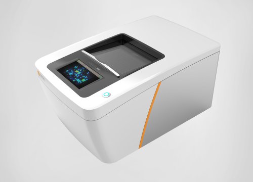
Amyotrophic lateral sclerosis (ALS) is characterized by progressive loss of upper and lower motor neurons. With limited treatment options, there is an urgent need for improved disease models to aid drug discovery and development efforts. Improved access to induced pluripotent stem cells (iPSCs) are providing researchers with better models. The sensitive functional assays of the Maestro MEA platform are enabling scientists to characterize ALS and other neurodegenerative diseases, both to gain a better understanding of pathogenic mechanisms as well as insight into efficacy of potential novel therapeutics. Learn how the Maestro can accelerate ALS research with these selected publications:
- Intrinsic Membrane Hyperexcitability of Amyotrophic Lateral Sclerosis Patient-Derived Motor Neurons
- Biosensing system for drug evaluation of amyotrophic lateral sclerosis based on muscle bundle and nano-biohybrid hydrogel composed of multiple motor neuron spheroids and carbon nanotubes
- Edaravone activates the GDNF/RET neurotrophic signaling pathway and protects mRNA-induced motor neurons from iPS cells
- ALS/FTD-Linked Mutation in FUS Suppresses Intra-axonal Protein Synthesis and Drives Disease Without Nuclear Loss-of-Function of FUS
Intrinsic Membrane Hyperexcitability of Amyotrophic Lateral Sclerosis Patient Derived Motor Neurons
Wainger BJ, Kiskinis E, et al. Cell Reports. (2014)
Research has shown many ALS patients demonstrate increased neural excitability, which may contribute to eventual cell death. Using iPSCs, researchers assess the intrinsic nature of this hyperexcitability in several ALS-associated genes and mutations, and evaluate potential therapeutic options.
Highlights:
>> iPSC-derived motor neurons carrying a variety of ALS-associated mutations display expected hyperexcitability, as seen in vivo.
>> MEA provides functional measurement of hyperexcitability and evaluation of drug efficacy.
>> Gene correction and treatment with potassium channel activators both reduce mutation-associated hyperexcitability.
Biosensing system for drug evaluation of amyotrophic lateral sclerosis based on muscle bundle and nano-biohybrid hydrogel composed of multiple motor neuron spheroids and carbon nanotubes
Ha T, Park S, et al. Chemical Engineering Journal. (2023)
Improved in vitro modeling of mature neuromuscular junction (NMJ) formation could benefit ALS drug discovery and development. Researchers develop a novel 3D NMJ biosensing system using motor neuron spheroids, carboxylated carbon nanotubes, and muscle tissue.
Highlights:
>> Motor neuron spheroids and carboxylated carbon nanotubes are combined in a 3D nano-biohybrid hydrogel.
>> MEA assays are used to characterize functional maturation of motor neuron spheroids.
>> Integration of biohybrid neural tissues and muscle allow for assessment of muscle recovery and contraction to evaluate potential therapeutics.
Edaravone activates the GDNF/RET neurotrophic signaling pathway and protects mRNA-induced motor neurons from iPS cells
Li Q, Feng Y, et al. Molecular Neurodegeneration. (2022)
iPSCs from ALS patient donors are frequently used to evaluate ALS therapeutics, but motor neuron differentiation protocols often take several weeks. Investigators present a rapid motor neuron differentiation protocol using synthetic mRNA.
Highlights:
>> Synthetic mRNAs encoding Ngn2 and Olig2 transcription factors rapidly and efficiently induce motor neuron differentiation.
>> Edaravone efficacy and capacity to provide neuroprotection from oxidative stress is evaluated via MEA assay.
>> Transcriptomics provide evidence that edaravone activates neurotrophic factor signaling pathways.
ALS/FTD-Linked Mutation in FUS Suppresses Intra-axonal Protein Synthesis and Drives Disease Without Nuclear Loss-of-Function of FUS
López-Erauskin J, Tadokoro T, et al. Neuron. (2018)
Mutations in the FUS gene are an identified cause of both ALS and frontotemporal dementia (FTD), but mechanisms underlying disease progression are unclear. Researchers use humanized FUS mice to model disease and gain insight into potential mechanisms causing pathophysiology.
Highlights:
>> Humanized mutant FUS mice exhibit age-dependent motor deficits, cognitive deficits, and synaptic loss, despite reduction in normal FUS function.
>> Primary mutant FUS neurons display synaptic dysfunction measured via MEA.
>> Pathophysiology is determined to be a result of a gain of toxicity due to accumulated FUS protein, stress response, and decreased synaptic function.


