Zabolocki M, McCormack K, van den Hurk M, Milky B, Shoubridge AP, Adams R, Jenne Tran J, Mahadevan-Jansen A, Reineck P, Thomas J, Hutchinson MR, Mak CKH, Añonuevo A, Chew LH, Hirst AJ, Lee VM, Knock E and Bardy C.
Nature Communications, 2020
Summary:
In this paper, the authors develop a neuromedium called BrainPhysTM Imaging (BPI) which optimized fluorescent and phototoxic compounds. Compared to available neuromedia, BPI enhances fluorescence signals, reduces phototoxicity and support electrical and synaptic activity of neurons in culture.
MEA and patch-clamp were used to determine the electrophysiological properties of human neurons in BPI. The network activity of human iPSC-derived neurons was compared in a 48-well multielectrode array (MEA) plate and did not show a significant difference in activity (synchrony, number of network events per minute, network interburst interval, mean network burst duration) with BPI compared to BP media. Cortical and midbrain human iPSC-derived neurons were matured for 52 days and spontaneous firing was recorded for 18 days (10 minute recording every 24 hours). The spontaneous firing decreased for the cultures with ACSF but was similar for BP and BPI.
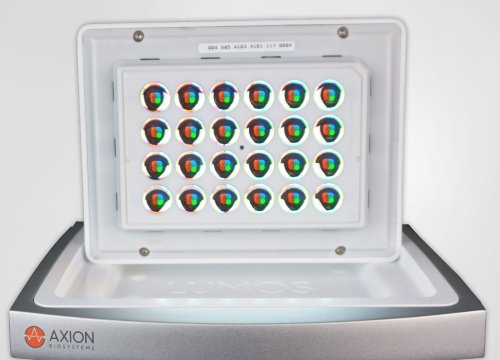
To test the long term effects of media with supplements for optogenetics experiments, the iPSC-derived neurons were evaluated in the Maestro MEA system. The neurons transfected with the optogene and stimulated with blue light (Lumos system) while on an MEA recorder were able to evoke action potentials with BPI.